Encyclopedia entry
Common Peroneal
Abstract
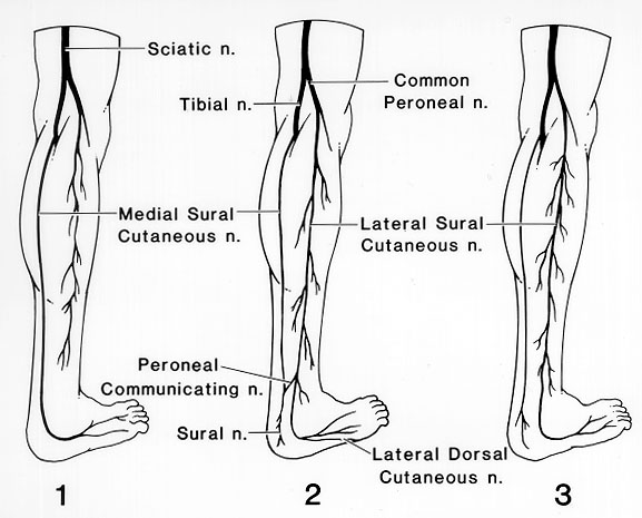
The sciatic usually divides into the common peroneal and tibial nerves at the level of the lower thigh. Very often these two nerves arise separately from the sacral plexus. They may be separated in the greater sciatic foramen by the piriformis muscle and pass into the thigh as contiguous but separate structures. Huelke (1958) reported that the peroneal communicating nerve arose directly from the common peroneal nerve in 54.7%, usually as a branch separate from the lateral sural cutaneous nerve (41.5%).
The sciatic usually divides into the common peroneal and tibial nerves at the level of the lower thigh. Very often these two nerves arise separately from the sacral plexus. They may be separated in the greater sciatic foramen by the piriformis muscle and pass into the thigh as contiguous but separate structures.
Huelke (1958) reported that the peroneal communicating nerve arose directly from the common peroneal nerve in 54.7%, usually as a branch separate from the lateral sural cutaneous nerve (41.5%).
The peroneal communicating nerve gave rise to the lateral sural cutaneous branches in 13.2% of sides studied (198 adult lower extremities). The peroneal communicating nerve was a terminal branch of the lateral sural cutaneous nerve in one-third of the sides, and arose from a trunk common to it and to the lateral sural cutaneous nerve in 12%.
The peroneal communicating nerve was absent in 19.7% of the 198 sides, and, due to its absence, no sural nerve was formed in these cases. When this occurs, it is usually the medial sural cutaneous nerve that passes onto the dorsum of the foot as the lateral dorsal cutaneous nerve. Only 58.6% of the cadavers had the same type of origin of the peroneal communicating nerve in both legs. No significant differences were found between the right and left sides, between sexes, or the place where the peroneal communicating or sural nerves arose. The union between the peroneal communicating and the medial sural cutaneous nerves was seen on 159 sides (80.3%). This union took place more often in the lower half of the leg.
Image 63A Image 63B Image 63C Image 63D Image 63E
Anatomy Atlases. Image hosted by the site it comes from. Open the original page.
Anatomy Atlases. Image hosted by the site it comes from. Open the original page.
Anatomy Atlases. Image hosted by the site it comes from. Open the original page.
Anatomy Atlases. Image hosted by the site it comes from. Open the original page.
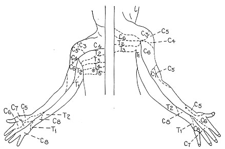
Anatomy Atlases. Image hosted by the site it comes from. Open the original page.
Image 63F Image 63G Image 63H Image 63I Image 63J
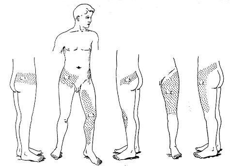
Anatomy Atlases. Image hosted by the site it comes from. Open the original page.
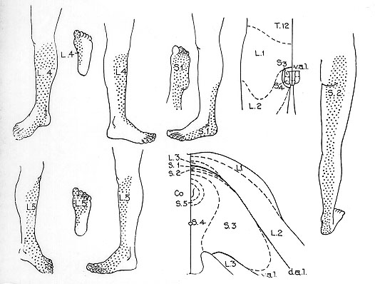
Anatomy Atlases. Image hosted by the site it comes from. Open the original page.
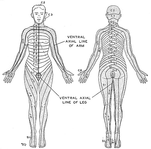
Anatomy Atlases. Image hosted by the site it comes from. Open the original page.
Anatomy Atlases. Image hosted by the site it comes from. Open the original page.
Anatomy Atlases. Image hosted by the site it comes from. Open the original page.
Anatomy Atlases. Image hosted by the site it comes from. Open the original page.
Riferimenti dell’opera originale
Elenco riportato come compare nella fonte, senza normalizzazione dei formati bibliografici.
- Beaton, L.E. and B.J. Anson. (1938) The relation of the sciatic nerve and of its subdivisions to the piriformis muscle. Anat. Rec. 70:1-5.
- Calori, L. (1880) Sull'alta divisione del nervo grande ischiatico considerata come differenza nazionale e sulle varietà del muscolo piriforme. Mem. R. Accad. Sci. Istituto di Bologna S. 4. 2:623-633.
- Cutore, G. (1902) La division du grand nerf sciatique chez l'homme. Arch. Ital. Biol. 37:491.
- Fredericq, H. (1908) Un cas de division complète du nerf grand sciatique accompagnée d'une duplicité complète du muscle pyramidal. Assoc. Anatomistes Comptes Rendus 10:168-170.
- Huelke, D.F. (1958) The origin o the peroneal communicationg nerve in adult man. Anat. Rec. 132: 81-92.
- Latarjet, A. (1948) Testut's Traite d'Anatomie Humain, 9th ed. G. Doin & Cie. Paris.
- Ming-tzu, P'an (1941) The relation of the sciatic nerve to the piriformis muscle in the Chinese. Am. J. Phy. Anthropol. 28:375-380.
- Misra, B.D. (1954) The relations of the sciatic nerve to the piriformis in Indian cadavers. J. Anat. Soc. India 3:44.
- Nizankowski, C., Slociak, J. and J. Szybejko. (1972) Variation of the course of the sciatic nerve in man. Folia Morphol. 31:451-456.
- Parsons, F.G. and A. Keith. (1896-97) Sixth annual report of the Committee of Collective Investigation of the Anatomical Society of Great Britian and Ireland. J. Anat. Physiol. 31:31-44.
- Schaefer, E.A., Symington, J. and T.H.Bryce., Eds. (1915) Quain's Anatomy, 11th ed., Longmans, Green, and Co., London.
- Trotter, M. (1932) The relation of the sciatic nerve to the piriformis muscle. Anat. Rec. 52:321-323.
Anatomical classification
- Systems:
- Nervous system
Content provenance
Adaptation
Adaptation of «Common Peroneal — Illustrated Encyclopedia of Human Anatomic Variation: Opus I: Muscular System» by Ronald A. Bergman, Adel K. Afifi, Ryosuke Miyauchi. Source: https://www.anatomyatlases.org/AnatomicVariants/NervousSystem/Text/CommonPeroneal.shtml Original work distributed under the CC BY-NC-SA 4.0 licence (https://creativecommons.org/licenses/by-nc-sa/4.0/deed.it). Adaptation by Redazione AIVA. Changes from the original: Testo originale inglese riprodotto integralmente e riorganizzato nel formato scheda del portale (sintesi, testo, riferimenti), senza modificarne le affermazioni. La traduzione italiana non è ancora stata eseguita, e la pagina lo dichiara. Le figure non sono state copiate: sono collegate alla pagina d’origine. This publication is not approved or endorsed by the authors of the original work.
Licence
This content is distributed under the licence CC BY-NC-SA 4.0. Licences and attribution