Encyclopedia entry
Coracobrachialis
Abstract
This muscle is chiefly a flexor muscle, although in the arm it corresponds to the adductor mass seen in the thigh. It develops from a tissue mass that forms biceps and brachialis and is supplied by the musculocutaneus nerve. The muscle actually consists of three parts: (a) a proximal part arising from the coracoid process and inserted onto the humerus close to the lesser tubercle; (b) a middle part of intermediate size; and (c) a distal part, which is the largest and most superficially placed.
This muscle is chiefly a flexor muscle, although in the arm it corresponds to the adductor mass seen in the thigh. It develops from a tissue mass that forms biceps and brachialis and is supplied by the musculocutaneus nerve.
The muscle actually consists of three parts: (a) a proximal part arising from the coracoid process and inserted onto the humerus close to the lesser tubercle; (b) a middle part of intermediate size; and (c) a distal part, which is the largest and most superficially placed. The muscle may extend to the medial epicondyle or near it, or it may be inserted into a supracondylar process.
In humans the middle part is the most constant, but it is generally accompanied by a part of the distal portion, with the musculocutaneus nerve passing between them. In 3.5-6.5% of arms the muscle is not traversed by the musculocutaneus nerve. The distal part is occasionally found quite separate from the other parts. The proximal part is represented occasionally by a proximal extension of insertion onto the surgical neck of the humerus or capsule of the shoulder joint, or by an accessory head (coracobrachialis superior or brevis) . The distal portion is sometimes present in the form of the coracobrachialis inferior or longus (Wood), and may be attached to the humerus, to a fibrous band of the medial intermuscular septum (ligament of Struthers), or even to the medial epicondyle.
Mori reported the separation of the belly into superficial and deep layers as follows: a. The belly of the muscle is completely separated into its component superficial and deep layers in 8 arms (16%). b. The belly of the muscle is incompletely separated into its component parts in 4 arms (8%). c. The belly shows no signs of dissociation into superficial or deep layers in 38 arms (76%).
In three (6%) instances, Mori found a deviant slip arising from the distal (deep) portion of coracobrachialis that ran medialward to fuse with the medial surface of the terminal portion of pectoralis major.
The muscle is sometimes connected to the brachialis muscle (Hyrtl, Wood) and a fasciculus has been observed joining the medial head of the triceps.
Coracobrachialis minor s. secundus (Gruber), coracobrachialis brevis s. rotator humeri (Wood), le court coracobrachialis (Cruveilhier) is an accessory muscle originating from the coracoid process; it crosses the radial nerve in the axilla and inserts into the tendinous part of latissimus dorsi.
Coracocapsularis (Wood) arises from the coracoid process and inserts into the shoulder capsule. Coracobrachialis may be absent.
Syn. : m. perforatus Casserii s. coracoideus, levator humeri internus (Arnold), Haken-arm muskel, Raben-Armmuskel (Hyrtl), Hakenmuskel.
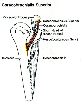
Coracobrachialis Superior
Redrawn and modified from Beattie — Anatomy Atlases. Image hosted by the site it comes from. Open the original page.
Riferimenti dell’opera originale
Elenco riportato come compare nella fonte, senza normalizzazione dei formati bibliografici.
- Beattie, P.H. (1947) Description of bilateral coracobrachialis muscle. Anat. Rec. 97:123-126.
- Becker, A.E. (1963) Variation of the M. coracobrachialis. Acta Morphol. Neer. Scand. 5:217-220.
- Bianchi, S. (1886) Varietà muscolari. Sperimentale (Firenze). 57:113-125.
- Curnow, J. (1873) Notes of some irregularities in muscles and nerves. J. Anat. Physiol. 7:304-309.
- Macalister, A. (1875) Additional observations on muscular anomalies in human anatomy (third series), with a catalogue of the principal muscular variations hitherto published. Trans. Roy. Irish Acad. Sci. 25:1-134.
- Mori, M. (1964) Statistics on the musculature of the Japanese. Okajimas Fol. Anat. Jap. 40:195-300.
- Odin, -. (1869) Faisceau supplémentaire du long chef du biceps et coraco-brachial surnuméraire sur le méme bras. Lyon Mêd. 2:1869.
- Reid, R.W. and S. Taylor. (1879) Anatomical variations. Reports. St. Thomas's Hospital 9:46.
- Wood, J. (1864) On some varieties in human myology. Proc. Roy. Soc. Lond. B 13: 299-303.
- Wood, J. (1867) On human muscular variations and their relation to comparative anatomy. J. Anat. Physiol. 1:44-59.
- Wood, J. (1868) Variations in human myology observed during the winter session of 1867-68 at King' College, London. Proc. Roy. Soc. Lond. B 17: 483-525.
- Zagorsky, P. (1809) Observationés anatomicae de musculorum quorundam corporis humani varietate minus frequente. Mem. d. L'Acad. Imp. d. Sci. d. St. Petersbourg T.1:355-359.
Anatomical classification
- Systems:
- Muscular system
Content provenance
Adaptation
Adaptation of «Coracobrachialis — Illustrated Encyclopedia of Human Anatomic Variation: Opus I: Muscular System» by Ronald A. Bergman, Adel K. Afifi, Ryosuke Miyauchi. Source: https://www.anatomyatlases.org/AnatomicVariants/MuscularSystem/Text/C/31Coracobrachialis.shtml Original work distributed under the CC BY-NC-SA 4.0 licence (https://creativecommons.org/licenses/by-nc-sa/4.0/deed.it). Adaptation by Redazione AIVA. Changes from the original: Testo originale inglese riprodotto integralmente e riorganizzato nel formato scheda del portale (sintesi, testo, riferimenti), senza modificarne le affermazioni. La traduzione italiana non è ancora stata eseguita, e la pagina lo dichiara. Le figure non sono state copiate: sono collegate alla pagina d’origine. This publication is not approved or endorsed by the authors of the original work.
Licence
This content is distributed under the licence CC BY-NC-SA 4.0. Licences and attribution