Encyclopedia entry
Digastricus (Anterior belly)
Abstract
The anterior belly of the digastric is highly variable. Mori , having studied 262 cadavers, classified the into 7 types as follows: A close relationship exists between the mylohyoid and the anterior belly of the digastric. There is usually some exchange of fibers sometimes amounting to complete fusion of the two muscles. Doubling of the anterior belly is a rather common variation, and the anterior bellies of opposite sides may be united by conversion of the fascia that typically passes between them into muscular tissue.
The anterior belly of the digastric is highly variable. Mori , having studied 262 cadavers, classified the into 7 types as follows:
- The "normal", in the anterior belly arises from the fossa digastricus of the manadible, and passes downward, backward and slightly outward to become continuous with the intermediate tendon (46.1% 0f 262 cadavers).
- The "ape" form, the anterior belly arises not only from the intermediate tendon of digastricus but also from the aponeurosis intertendiones (aponeurosis interdiagastricque [Zlabek]). The muscle fibers arise from the intertendinous aponeurosis and intermediate tendon and insert on the inner surface of the mandible (5 of 262 cadavers).
- The anterior type. The muscle fibers of this type arise from the aponeurosis intertendines and form the muscle plate, and this plate fuses with the muscle fibers arising from the intermediate tendon of digastricus to make one broad plate. Mori finds two forms of this type; a. the continous form where the muscle arising from the aponeurosis intertendines and the anterior belly of digastricus fuse (found bilaterally in 10.3% of 262 cadavers, and unilaterally in 6.8% (right and left side equally) of 262 cadavers and b. the discontinuous form where there is a boundry between the muscle arising from the intertendinus aponeurosis and those arising from the anterior belly of digastricus (bilaterally) in 0.3% of 262 cadavers, and on one side in 1.9% (3 right, 2 left) of 262 cadavers.
- The posterior type, where the muscle fibers arising from the aponeurosis intertendines makes a thin muscle plate and inserts onto the mandible. The boundry between the anterior belly of digastricus and the unusual muscle plate is clearly defined. There are two forms of this type according to Mori. a) the continous form where the variant muscle plate passes anteriorly and inserts onto the inner surface of the mandible. Some of these muscle fibers join mylohyoideus. On both the right and left sides the frequency of this variation is 0.7%. b) In the myloid form the deviant muscle fibers pass medially and fuse with the mylohyoid o the opposite side. If the variant is bilateral the deviant muscle plate has a raphe in the median line.
- The biceps form where the anterior belly is divided into two heads, the medial head goes to the opposite side to be inserted into the fossa digastricus of the opposite side. The insertion of the variant muscle is always superior to that of the anterior belly.
- Accessory muscle fascicles located between the mandible and the anterior belly of digastricus is very rarely seen. The deviant muscle fibers arise from the lateral margin of the anterior belly of the digastric and passes to the mandible where it inserts. the thickness of the muscle belly also varies.
- The combination form is composed of elements of the preceeding six types and is very complicated. See illlustrations (e.g., those of Mori) of these complicated forms of the anterior belly of the diagastric muscle.
A close relationship exists between the mylohyoid and the anterior belly of the digastric. There is usually some exchange of fibers sometimes amounting to complete fusion of the two muscles. Doubling of the anterior belly is a rather common variation, and the anterior bellies of opposite sides may be united by conversion of the fascia that typically passes between them into muscular tissue. An independent muscle, mentohyoid (Macalister), extending between the body of the hyoid and the symphysis of the mandible, may be found along the medial border of the anterior belly, and may be considered as a separate part of the digastric. The anterior belly may be absent. Tri- and quadrigastric muscles have been reported.
The variability of the anterior belly of the digastric muscle can be seen in the following listing provided by Mori from Loth. Atypical anterior bellies were found in 6.6% of cases by Hallett (1847), in 5.9% by Wood (1862), in 6.0% by LeDouble (1891), in 12% by Loth, in 26% by Bavero (1896), in 36% (fetal) by Bianchi (1896), in 48% by Stracher (1908), in 51% by Bianchi (1890), in 52% by Ruffini (1908), in 52.7% by Stracker (1928), in 63.3% by Zalbek (1933) and in 73% by Bertalli (1927). The anterior belly of digastricus is a branchiomeric muscle derived from the mesoderm associated with the branchial arches. It is innervated by the trigeminal (mandibular division) nerve.
Syn.: m. biventer mandibulae, biventer, digastricus maxillae inferioris, digastricus ossis hyoidei (M.J. Weber), Zweibauchiger Unterkiefermuskel, digastrique.
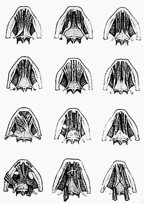
Varieties of the Anterior Belly of Digastricus.
from Ruffini, 1908 — Anatomy Atlases. Image hosted by the site it comes from. Open the original page.
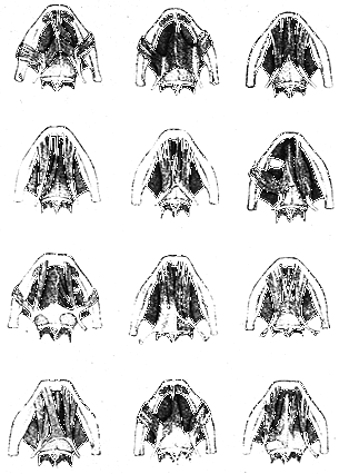
Varieties of the Anterior Belly of Digastricus.
from Ruffini, 1908 — Anatomy Atlases. Image hosted by the site it comes from. Open the original page.
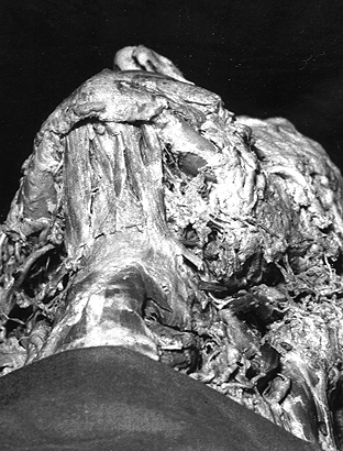
Anterior Belly of Digastricus
- Laboratory specimen, 1993.
University of Iowa. — Anatomy Atlases. Image hosted by the site it comes from. Open the original page.
References
Bovero, A. (1896) Sur les muscles diagastriques de l'os hyoïde. Arch. Ital. Biol. 26:158.
Corsy, F. (1920) Un nouveau cas d'appareil hyoïdien. Anomalies multiples des muscles du deuxième arc. Soc. Biol., Comptes Rendus Hebdomadaires des Seances et Memoires. 83:366-368.
Dratsch, S. (1930) Ein Fall von variationen des M. digastricus mandibulae. Anat. Anz. 69:81-82.
Fleishmann, G. (1810) Anatomische Wahrnehmungen. Abhandlungen der Physikalish-Medizinischen Sozieta zu Erlangen. 1:23-36.
Focacci, M. (1901) Contribution a l'étude du muscle interdiagastrique de Bianchi. Arch. Ital. Biol. 36;476.
Gruber, W. (1880 a) Über den Musculus trigastricus maxillae inferioris. Arch. Path. Anat. Physiol. Klin. Med. 81:445-449.
Gruber, W. (1880 b) Musculus digastricus maxillae inferioris mit Ursprung seines vorderen Bäuches an und hinter der Mitte des Seitentheiles der Maxilla im Bereiche der Strecke zwischen dem Ansatze des masseter und dem Ursprung des M. depressor anguli oris. Arch. Path. Anat. Physiol. Klin. Med. 81:449-453.
Gruber, W. (1885) Auftreten der Bäuche des Musculus digastricus maxillae inferioris als selbständige Muskeln Musculus mento-hyoideus et mastoideo-maxillaris. Arch. Path. Anat. Physiol. Klin. Med. 101:253-255.
Hahn, H. (1910) Eine seltene Anomalie des vordern Bauches des M. digastricus mandibulae. Zeitschrift für Morphologie und Anthropologie. 13:281-288.
Macalister, A. (1875) Additional observations on muscular anomalies in human anatomy (third series), with a catalogue of the principal muscular variations hitherto published. Trans. Roy. Irish Acad. Sci. 25:1-134.
Mori, M. (1964) Statistics on the musculature of the Japanese. Okajimas Fol. Anat. Jap. 40:195-300.
Ruffini, A. (1908) Sul muscolo interdigastrico di Bianchi e sull'aponeurosi soprajoidea od intermediojoidea. Rarissimo caso di mancanza bilaterale del ventre anteriore del m. digastrico della mandibola. Arch. Ital. Anat. e di Embriol. 7:578-601.
Tanaka, M., Onda, S., and T. Tsukamoto. (1958) The situation of the hyoidal muscles (M. digastricus mandibulae). Kaibogaku Zasshi. 33:Supple., 113-114. In Japanese.
Tojima, T. and T. Nakano. (1950) Study on the venter anterior of the M. digastricus mandibulae. Iwate Daigaku Kaibogakukyoshitsu Gyosekishu. 1:111-123. In Japanese.
Tsukamoto, T. and S. Tanaka. (1958) On the digastricus mandibulae, M. stylohyoideus. Tokyo Shikadaigaku Kaibogakukyoshitsu Gyosekishu. 8:1-4. In Japanese.
Yamada, F. (1933) The M. digastricus mandibulae of the fetus and new-born. Keio Igaku Zasshi 13:1503-1507. In Japanese.
Yamada, F. (1935) Observation on the venter anterior of the diagastricus of the adults and fetus. Kaibogaku Zasshi. 8:303-308. In Japanese.
Anatomical classification
- Systems:
- Muscular system
Content provenance
Adaptation
Adaptation of «Digastricus (Anterior belly) — Illustrated Encyclopedia of Human Anatomic Variation: Opus I: Muscular System» by Ronald A. Bergman, Adel K. Afifi, Ryosuke Miyauchi. Source: https://www.anatomyatlases.org/AnatomicVariants/MuscularSystem/Text/D/05Digastricus.shtml Original work distributed under the CC BY-NC-SA 4.0 licence (https://creativecommons.org/licenses/by-nc-sa/4.0/deed.it). Adaptation by Redazione AIVA. Changes from the original: Testo originale inglese riprodotto integralmente e riorganizzato nel formato scheda del portale (sintesi, testo, riferimenti), senza modificarne le affermazioni. La traduzione italiana non è ancora stata eseguita, e la pagina lo dichiara. Le figure non sono state copiate: sono collegate alla pagina d’origine. This publication is not approved or endorsed by the authors of the original work.
Licence
This content is distributed under the licence CC BY-NC-SA 4.0. Licences and attribution