Encyclopedia entry
Ethmoid Bone
Abstract
Ethmoidal variations include reduction in the size of lamina orbitalis, which occurs when the frontal or maxillary bones or both participate in the formation of the medial wall of the orbit. The orbital plate may be divided into two, three, or four parts by vertical fissures. Occasionally, the anterosuperior or anteroinferior angles are separated from the orbital plate forming ossicles termed ossiculum ethmolacrimale superius and ossiculum ethmolacrimal inferius. An increase in the number of nasal conchae is common.
Ethmoidal variations include reduction in the size of lamina orbitalis, which occurs when the frontal or maxillary bones or both participate in the formation of the medial wall of the orbit. The orbital plate may be divided into two, three, or four parts by vertical fissures. Occasionally, the anterosuperior or anteroinferior angles are separated from the orbital plate forming ossicles termed ossiculum ethmolacrimale superius and ossiculum ethmolacrimal inferius.
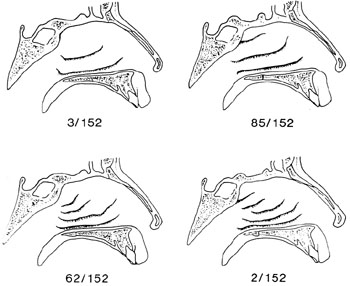
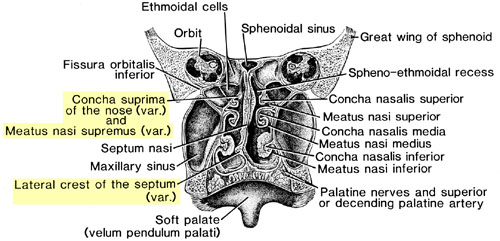
An increase in the number of nasal conchae is common. The ethmoid may exhibit two to four conchae (not including the inferior or maxilloturbinate). A bony process below the middle concha may descend over the aperture of the maxillary sinus. A fourth conchae over the superior one is called concha Santorini.
The anterior ethmoidal foramen in the lamina cribrosa may lead into a bony canal through which an artery and nerve course.
The crista galli may be pneumatized.
The lamina papyracea that covers the labyrinth laterally may exist as two bones resulting from a perpendicular suture. The posterior ethmoidal canal is frequently absent, being replaced by a canal at the junction between the upper edge of the lamina and the sphenoid body.
Secondary foramina of the lamina orbitalis are frequent in the aged. Reduction of this plate is found where either the maxilla or the frontal (or both) sends processes to participate in forming the medial wall of the orbit.
The textbook number of ethmoid foramina is two; Lang (1983) found them on the left side in 68.3% of skulls and on the right side in 67.4%. In the remainder there were 3 to 5, there were 3 foramina in about 40% of skulls.
Variation in Number of Meatuses in Nasal Fossae
Anatomy Atlases. Image hosted by the site it comes from. Open the original page.
Cavum Nasi - The Nasal Fossae
Anatomy Atlases. Image hosted by the site it comes from. Open the original page.
Riferimenti dell’opera originale
Elenco riportato come compare nella fonte, senza normalizzazione dei formati bibliografici.
- Anson, B.J., Ed. (1966) Morris' Human Anatomy, 12th ed., The Blakiston Division, McGraw Hill Book Co., New York.
- Bianchi, S. (1985) Sur la division de "l'os Planum" de l'Ethmoïde dans le crâne de l'homme et des antropoïdes et sur la non-existence de l'os lacrymal postérieur chez quelques mammifères. Arch. Ital. Biol. 24:310-311.
- Fusari, R. (1897) Sur les différents modes de substitution de la partie postérieur de la lame papyraceé l'orbite de l'homme. Arch. Ital. Biol. 28:470-471.
- Gruber, W. (1875) Die Crista galli des Os ethmoideum mit einer Höhle. Arch. Pathol. Anat. Physiol. Klin. Med. 65:11.
- Lang, J. (1983) Clinical Anatomy of the Head, Neurocranium, Orbit and Craniocervical Region. Springer Verlag, Berlin, Heidelberg and New York.
- Menzel, K.M. (1934) über das Vorkommen von überzahligen muschelartigen Gebilden im hinteren Teil des mittleren Nasenganges. Anat. Anz. 78:229-240.
- Perovic, D. (1939) über eine ungewöhnliche Insertion der unteren Nasenmuschel. Anat. Anz. 88:133-139.
- Stieda, L. (1891) Uber den Sulcus ethmoidalis der Lamina cribrosa des Siebbeines. Anat. Anz. 6:232-237.
- Teatini, G., Simonetti, G., Salvoline, U., Masala, W., Meloni, F., Rovasio, S. and G.L. Dedola. (1987) Computed tomography of the ethmoid labyrinth and adjacent structures. Annals of Otology, Rhinology & Laryngology (Annals of Head & Neck Medicine & Surgery) 96(3):239-246.
Anatomical classification
- Systems:
- Skeletal system
Content provenance
Translation
Italian translation of «Ethmoid Bone — Illustrated Encyclopedia of Human Anatomic Variation: Opus I: Muscular System» by Ronald A. Bergman, Adel K. Afifi, Ryosuke Miyauchi. Source: https://www.anatomyatlases.org/AnatomicVariants/SkeletalSystem/Text/EthmoidBone.shtml Original work distributed under the CC BY-NC-SA 4.0 licence (https://creativecommons.org/licenses/by-nc-sa/4.0/deed.it). Translation by Redazione AIVA. Changes from the original: Traduzione italiana del testo originale inglese, che resta consultabile nella versione inglese di questa pagina. Il contenuto è stato riorganizzato nel formato scheda del portale (sintesi, testo, riferimenti) senza modificarne le affermazioni. Le figure non sono state copiate: sono collegate alla pagina d’origine. This publication is not approved or endorsed by the authors of the original work.
Licence
This content is distributed under the licence CC BY-NC-SA 4.0. Licences and attribution
