Encyclopedia entry
Extensor Pollicis Longus
Abstract
Doubling of this muscle is frequent, and the ulnar portion may pass beneath the dorsal ligament with the common extensor. It has been reported the this muscle is absent in about 1.5% of individuals. Macalister reported the variations of this muscle as follows: 1. It may receive a slip of extensor digitorum communis which runs in the same in the radius (wood); 2. Macalister found the muscle doubled with both tendons inserting as usual; 3. Giving a slip to the base of the first phalanx in a case where extensor pollicis longus was absent; 4.
Doubling of this muscle is frequent, and the ulnar portion may pass beneath the dorsal ligament with the common extensor. It has been reported the this muscle is absent in about 1.5% of individuals.
Macalister reported the variations of this muscle as follows:
1. It may receive a slip of extensor digitorum communis which runs in the same in the radius (wood); 2. Macalister found the muscle doubled with both tendons inserting as usual; 3. Giving a slip to the base of the first phalanx in a case where extensor pollicis longus was absent; 4. A slip from the extensor pollicis longus to the first phalanx coexisting with a slip from extensor pollicis brevis to the second phalanx.
A slip from the tendon of the long extensor to extensor indicis is seen occasionally. A rarer variation is an additional extensor between extensor indicis and extensor pollicis longus, with a doubled tendon and insertion onto both digits. See paragraph on extensor indicis proprius.This additional extensor may replace extensor pollicis longus or extensor indicis.
Extensor pollicis et indicisis a remarkable muscle occasionally found, and whose homologue is common among other groups of animals including the dog, fox, wolf, jackel, panther, and the dingo; it has been described by Wood, Clason and by Macalister. Its most common arrangement is an origin fron the ulna between the extensor indicis and extensor pollicis longus (with which it usually coexists); it usually is also connected to the interosseous ligament and to the intermuscular septa; its tendon passes downward and divides into two, one to the thumb, and the other to the index finger; sometimes the former runs into the indicial tendon of the extensor communis, while the latter passes to the first phalanx of the thumb, or one is inserted into the first phalanx of the index finger (Wood), or one has been seen joing the extensor pollicis brevis tendon, or substitute for it, or inserted into the second and third phalanges of the index finger, or into the common extensor aponeurosis of the index, or with the extensor pollicis longus. It has also been found arising from the intermuscular septum between the extensor digitorum communis and the extensor pollicis longus, forming a layer intermediate between them, and at first sight apparently connected with the common extensor; this case was inserted, with the tendon of the extensor of the distal phalanx of the thumb; and with the lateral of the two tendons of a doubled indicator. Abductor pollicis tertius (extensor atque abductor pollicis accessorius), a rare muscle, arises from the dorsal aspect of the radius with abductor pollicis longus and inserts, after fusion with abductor pollicis brevis, onto the first metacarpal.
Syn.: m. exttensor. pollicis. major, extensor. secundi internodii pollicis.
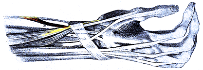
Note the muscular slip (red) that extends from the tendon of brachioradialis to the tendon (yellow) of abductor pollicis longus
From Calori. — Anatomy Atlases. Image hosted by the site it comes from. Open the original page.
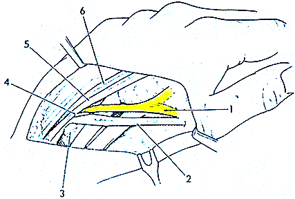
Extensor pollicis et indicis .
from Calori. — Anatomy Atlases. Image hosted by the site it comes from. Open the original page.
Riferimenti dell’opera originale
Elenco riportato come compare nella fonte, senza normalizzazione dei formati bibliografici.
- Anson, B.J., Ed. (1966) Morris' Human Anatomy. 12th ed. The Blakiston Division, McGraw-Hill Book Company, New York.
- Baumann, J.A. (1947) Valeur, variation, et équivalences des muscles extenseurs, interosseux, adducteurs et abducteurs de la main et du pied chez l'homme. Acta Anat. 4:10-16.
- Breglia, A. (1886) Di una anomalia del plesso brachiale. Rivista Internazionale di Medicina e Chirurgia. 4:337-344.
- Calori, L. (1867) Di alcune varieta muscolari dell'avambraccio e dell'eminenza ipothenar. Mem. Accad. R. Sci. Istituto di Bologna. S.2. 7:359-381.
- Debierre, C. (1888) Anomalie des muscles radiaux externes et du long abductor du pouce de l'homme. Soc. Biol. Comptes Rendus Hebdomadaires des Séances et Mémoires. 40:547-550.
- Dubreuil-Chambardel, L. (1919) Des muscles fléchisseurs et extenseurs sur un pouce a trois phalanges. Bull. Mem. Soc. d'Anthropol. Paris. 10:45-47.
- Gruber, W. (1881) Über den dem constanten Musculus extensor pollicis et indicis gewisser Säugetiere homologen supernumerären Muskel beim Menschen. Arch. Path. Anat. Physiol. Klin. Med. 86:471-491.
- Gruber, W. (1882) Über den zum Extensor pollicis et indicis singularis gewordenen extensor pollicis longus beim Menschen. Arch. Path. Anat. Physiol. Klin. Med. 90:103-108.
- Gruber, W. (1885) Über die im Bereiche des Lig. carpi dorsale schon congenital und in der Norm vorkommende Communication der Sehenscheide für den Extensor pollicis longus mit der für die Radialis externi (nach 500 untersuchungen), und über einen Fall von Hygroma proliferum dieser mit einander communicirender Sehnenscheiden. Arch. Path. Anat. Physiol. Klin. Med. 101:245-253.
- Gruber, W. (1885) Bcobachtung eines Schnenrisses des Musculus extensor pollicis longus in 3 Hillen. Archiv f. Pathologische Anatomic u. Physiologic u. f. klinische Medicin 102(l):1-2.
- Henle, J. (1871) Handbuch der Muskellehre des Menschen, in Handbuch der systematischen Anatomie des Menschen. Verlag von Friedrich Vieweg und Sohn, Braunschweig.
- Kaplan, E.B. and P. Nathan. (1969) Accessory extensor pollicis longus. Bull. NY Hosp. Joint Dis. 30:202-207.
- Kawaai, M. (1935) Accessory tendon from the terminal tendon of the M. extensor pollicis longus. Kaibogaku Zasshi 8:261-275. In Japanese.
- Krishnamurti, A. (1962) Anomalous insertion of extensor pollicis longus. J. Anat. Soc. India 11:90-91.
- LeDouble, A.F. (1897) Traité des Variations du Système Musculaire de l'Homme et leur Signification au Point de Vue de l'Anthropology Zoologique., Librairie C. Reinwald, Schleicher Freres Eds., Paris.
- Macalister, A. (1875) Additional observations on muscular anomalies in human anatomy (third series), with a catalogue of the principal muscular variations hitherto published. Trans. Roy. Irish Acad. Sci. 25:1-134.
- Reid, R.W. and S. Taylor. (1879) Anatomical variations. Reports. St. Thomas's Hospital 9:48.
- Salama, R. and S.L. Weissman. (1975) Congenital bilateral anomalous band between flexor and extensor pollicis longus tendons. Hand 7:25-26.
- Shepherd, F.J. (1880) Notes of abnormalities observed in the dissecting-room of the McGill University from October 1875, to May, 1879. Ann. Report: Montreal General Hospital 1:71-93.
- Straus, Jr., W.L. and O.Temkin. (1943) Vesalius and the problem of variability. Bull. History Med. 14:609-633.
- de Vilhena, H. (1933) Le muscle surnuméraire extenseur commun du pouce et de l'index. Assoc. Anat., Comptes Rendus 28:685-702.
- Wood, J. (1868) Variations in human myology observed during the winter session of 1867-68 at King's College, London. Proc. Roy. Soc. Lond. B 16:483-525.
Anatomical classification
- Systems:
- Muscular system
Content provenance
Adaptation
Adaptation of «Extensor Pollicis Longus — Illustrated Encyclopedia of Human Anatomic Variation: Opus I: Muscular System» by Ronald A. Bergman, Adel K. Afifi, Ryosuke Miyauchi. Source: https://www.anatomyatlases.org/AnatomicVariants/MuscularSystem/Text/E/27Extensor.shtml Original work distributed under the CC BY-NC-SA 4.0 licence (https://creativecommons.org/licenses/by-nc-sa/4.0/deed.it). Adaptation by Redazione AIVA. Changes from the original: Testo originale inglese riprodotto integralmente e riorganizzato nel formato scheda del portale (sintesi, testo, riferimenti), senza modificarne le affermazioni. La traduzione italiana non è ancora stata eseguita, e la pagina lo dichiara. Le figure non sono state copiate: sono collegate alla pagina d’origine. This publication is not approved or endorsed by the authors of the original work.
Licence
This content is distributed under the licence CC BY-NC-SA 4.0. Licences and attribution