Encyclopedia entry
Platysma
Abstract
There is usually decussation of the two contralateral muscles, across the midline, especially in their upper parts, where a certain amount of such crossing can be considered typical. The muscles development is subject to considerable variation, sometimes forming a very thin, pale layer largely interspersed among connective tissue, and at other times forming strong, deep red muscle bundles with much less intermixture of connective tissue (collagen). Its extension upon the face may also vary considerably, sometimes being traceable as high as the zygoma and, dorsally, to behind the ear.
There is usually decussation of the two contralateral muscles, across the midline, especially in their upper parts, where a certain amount of such crossing can be considered typical. The muscles development is subject to considerable variation, sometimes forming a very thin, pale layer largely interspersed among connective tissue, and at other times forming strong, deep red muscle bundles with much less intermixture of connective tissue (collagen). Its extension upon the face may also vary considerably, sometimes being traceable as high as the zygoma and, dorsally, to behind the ear. On the other hand, the muscle may be considerably reduced in size, especially distally; complete absence of the lower half has been reported. Rarely, a deep transverse layer is found. Platysma is intimately but variably associated with mentalis.
A rare case of a muscle described as a sphincter colli profundus has been reported. This muscle sheet arose deep to platysma from the medial part of the clavicle and inserted into the fascia of the pinna. In this particular case, platysma was reduced in size and supraclavicularis and mandibulomarginalis muscles were present.
Syn.: m. subcutaneus colli, platysma myoides, quadratus genae (Cowper), latissimus colli (Albin), Breiter Halsmuskel, Hauthalsnmuskel, Le peaucier (Winslow).
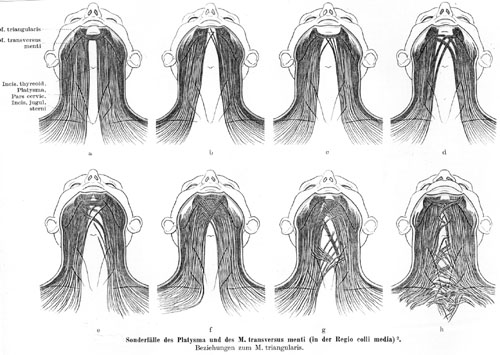
Platysma, 24 Varieties
Anatomy Atlases. Image hosted by the site it comes from. Open the original page.
Riferimenti dell’opera originale
Elenco riportato come compare nella fonte, senza normalizzazione dei formati bibliografici.
- Anson, B.J.,Ed. (1966) Morris' Human Anatomy. The Blakiston Division, McGraw-Hill Book Company, New York.
- Huber, E. (1918-19) des Sphincter colli profundus beim Menschen. Anat. Anz. 51:480-492.
- Kobayashi, T. (1948) On the platysma of Japanese in the HOKURIKU district. Juzenkai Igaku Zasshi 51:129. In Japanese.
- Macalister, A. (1875) Observations on muscular anomalies in the human anatomy. Third series with a catalogue of the principal muscular variations hitherto published. Trans. Roy. Irish Acad. Sci. 25:1-130.
- Morita, M. (1943). On the platysma of the Japanese and Corean. Kaibogaku Zasshi 21:755-767. In Japanese.
- Onda, S. and Y. Kobayashi. (1958) Morphological studies of the facial portion of the platysma. Shika Gakuho 58: Supple. 1-8. In Japanese.
- Sanchez, -. and M. Sanchez. Quelques variations morphologiques des muscles peauciers de la tê. L'Assoc. Anatomistes, Comptes rendus 25:586-582.
- Shima, H. (1959) Anatomical studies on the M. platysma. Shika Gakuho 59:261.
- Taketa, R., Takeishi, M., and T. Esaki. (1955) On the M. platysma. Kurume Igakkai Zasshi 18:379-384. In Japanese.
- von Lanz and W. Wachsmuth. (1955) Prakische Anatomie. Springer-Verlag; Berlin.
Anatomical classification
- Systems:
- Muscular system
Content provenance
Translation
Italian translation of «Platysma — Illustrated Encyclopedia of Human Anatomic Variation: Opus I: Muscular System» by Ronald A. Bergman, Adel K. Afifi, Ryosuke Miyauchi. Source: https://www.anatomyatlases.org/AnatomicVariants/MuscularSystem/Text/P/30Platysma.shtml Original work distributed under the CC BY-NC-SA 4.0 licence (https://creativecommons.org/licenses/by-nc-sa/4.0/deed.it). Translation by Redazione AIVA. Changes from the original: Traduzione italiana del testo originale inglese, che resta consultabile nella versione inglese di questa pagina. Il contenuto è stato riorganizzato nel formato scheda del portale (sintesi, testo, riferimenti) senza modificarne le affermazioni. Le figure non sono state copiate: sono collegate alla pagina d’origine. This publication is not approved or endorsed by the authors of the original work.
Licence
This content is distributed under the licence CC BY-NC-SA 4.0. Licences and attribution