Encyclopedia entry
Rhomboideus Major, Rhomboideus Minor
Abstract
The rhomboid sheet may extend beyond its usual limits in both a cranial and caudal direction. In some cases, the caudal border may reach the cranial border of latissimus dorsi. The caudal extension of rhomboideus major may be simultaneously associated with a cranial extension of latissimus dorsi. Mori reported the upper limit of the vertebral origin to be as follows: Upper Limit: Cervical 4 right side, 5 left side, 5 Total 10 16.7%. 5 right side, 18 left side, 18 Total 36 60.0%. 6 right side, 7 left side, 7 Total 14 23.3%. Lower Limit: Thoracic 3 right side, 1 left side,1 Total 2 9.3%.
The rhomboid sheet may extend beyond its usual limits in both a cranial and caudal direction. In some cases, the caudal border may reach the cranial border of latissimus dorsi. The caudal extension of rhomboideus major may be simultaneously associated with a cranial extension of latissimus dorsi.
Mori reported the upper limit of the vertebral origin to be as follows:
Upper Limit: Cervical 4 right side, 5 left side, 5 Total 10 16.7%. 5 right side, 18 left side, 18 Total 36 60.0%. 6 right side, 7 left side, 7 Total 14 23.3%.
Lower Limit: Thoracic 3 right side, 1 left side,1 Total 2 9.3%. 4 right side, 10 left side, 10 Total 20 33.3%. 5 right side, 14 left side, 14 Total 28 46.6%. 6 right side, 5 left side, 5 Total 10 16.6%. Length of Origin: C4-T3 (7 vertebrae) right side, 1 left side, 1. C4-T4 (8 vertebrae) right side, 3 left side, 3. C5-T4 (7 vertebrae) right side, 7 left side, 7. C5-T5 (8 vertebrae) right side, 11 left side, 11. C6-T5 (7 vertebrae) right side, 3 left side, 3. C6-T6 (8 vertebrae) right side, 5 left side, 5.
Note that the length of origin is most frequently 8 spinous processes.
Rhomboideus minor may be divided into separate bundles; the same may occur for the major muscle. A deep lamella may be present , taking the form of a slip of muscle fibers arising from the cranial part of the sheet but crossing obliquely from the main mass of the fibers to reach the inferior angle of the scapula. Slips from the tendinous insertion of the muscle may sweep over and into the fascia of infraspinatus, or into teres major, or the scapular origin of latissimus dorsi.
An additional muscle has been reported extending close to the upper border of the minor muscle, from the scapula to the occipal bone, and has been named rhomboideus-occipitalis or capitis (occipitoscapularis [Wood]) .
Rhomboideus-occipitalis passes between trapezius and splenius to reach the occipital bone.
In a modified form this accessory muscle may be present as a slip arising form the transverse process of the atlas or, the fascia over splenius capitis and inserting onto the vertebral border of the scapula, or onto the surface of serratus posterior superior. It is believed that the descent of the scapula in ontogeny leads to the characteristic obliquity of the rhomboids.
Rhomboideus minor has been reported absent more frequently than rhomboideus major. These two muscles may be inseparable and appear a a single muscle or they may be divided in several fascicles making their identification as discrete entities difficult.
A few additional slips of muscle have been identified associated with the rhomboids; 1) slips to latissimus dorsi and to teres major. 2) a slip at the level of rhomboideus minor, named rhomboideus minimus, extends from the scapula to thoracic or lower cervical vertebral spines.
Balli has provided the following details regarding the insertion of the rhomboids to the scapula. In 54% of cases, the upper and middle fibers are attached by weak tendinous fibers to the vertebral border below rhomboideus minor, the lower fibers by a strong tendon near to the inferior angle. In 21% of cases the insertion is by a fibrous arch attached weakly above to the vertebral border below rhomboideus minor, but strongly below on the inferior angle, where it receives the primary mass of muscle fibers. In some instances (6%) the arch is replaced by two or three smaller arches. In 19% of cases, all the fibers converge on the inferior angle so that the muscle is triangular, not rhomboidal.
Syn.: m. Rma; rhomboideus inferior, Grosser Rautenmuskel.
m. RMi; rhomboideus superior, Kleiner Rautenmuskel.
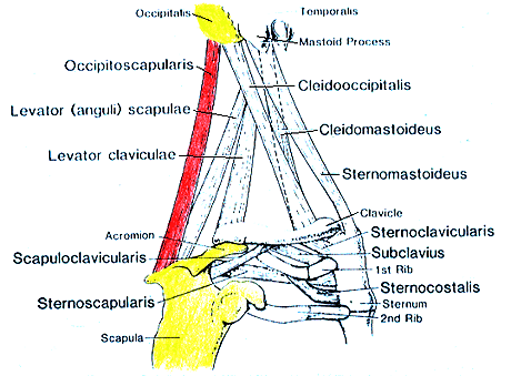
Varieties of Chest, Neck, and Shoulder Muscles-Occipitoscapularis.
- modified and redrawn from Wood.
Anatomy Atlases. Image hosted by the site it comes from. Open the original page.
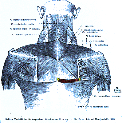
Rhomboideus Minimus.
from Haffner. — Anatomy Atlases. Image hosted by the site it comes from. Open the original page.
Riferimenti dell’opera originale
Elenco riportato come compare nella fonte, senza normalizzazione dei formati bibliografici.
- Anson, B.J., Ed. (1966) Morris' Human Anatomy,12th ed. The Blakiston Division, McGraw-Hill Book Company, New York.
- von Haffner, H. (1903) Eine Seltene doppelseitige Anomalie des Trapezius. Internat. Monatsschrift für Anat. Physiol. 20:313-318.
- Henle, J. (1871) Handbuch der Muskellehre des Menschen, in Handbuch der systematischen Anatomy des Menschen. Verlag von Friedrich Vieweg und Sohn, Braunschweig.
- Kitano, S., Takata, N. and M. Sato. (1957) One case of the variation of the M. rhomboideus minor. Kurume Igakkai Zasshi. 20:1045-1048. In Japanese.
- LeDouble, A.F. (1897) Traitédes Variations du Systeme Musculaire de l'Homme et Leur Signification au Point de vue de l'Anthropologie Zoologique. Libraire C. Reinwald, Scheicher Freres, Paris.
- Macalister, A. (1875) Observations on muscular anomalies in the human anatomy. Third series with a catalogue of muscular variations hitherto published. Trans. Roy. Irish Acad. Sci. 25:1-130.
- Mori, M. (1964) Statistics on the musculature of the Japanese. Okajimas Folia Anatomica Japonica 40:195-300.
- Mori, -. and -. Minoru. (1950) In the M. rhomboideus. Sotai Taiji no Kaibogakuteki Kenkyu 11:38-78. In Japanese.
- Schaefer, E.A., Symington, J. and T.H. Bryce., Eds. (1923) Quain's Anatomy, 11th ed. Longmans, Green and Co., London.
- Selden, B.R. (1935) Congenital absence of trapezius and rhomboideus major muscles. J. Bone Joint Surg. 171058-1059.
- Wood, J. (1867) On human muscular variations in their relation to comparative anatomy. J. Anat. Physiol. 1:44-59.
Anatomical classification
- Systems:
- Muscular system
Content provenance
Adaptation
Adaptation of «Rhomboideus Major, Rhomboideus Minor — Illustrated Encyclopedia of Human Anatomic Variation: Opus I: Muscular System» by Ronald A. Bergman, Adel K. Afifi, Ryosuke Miyauchi. Source: https://www.anatomyatlases.org/AnatomicVariants/MuscularSystem/Text/R/23Rhomboideus.shtml Original work distributed under the CC BY-NC-SA 4.0 licence (https://creativecommons.org/licenses/by-nc-sa/4.0/deed.it). Adaptation by Redazione AIVA. Changes from the original: Testo originale inglese riprodotto integralmente e riorganizzato nel formato scheda del portale (sintesi, testo, riferimenti), senza modificarne le affermazioni. La traduzione italiana non è ancora stata eseguita, e la pagina lo dichiara. Le figure non sono state copiate: sono collegate alla pagina d’origine. This publication is not approved or endorsed by the authors of the original work.
Licence
This content is distributed under the licence CC BY-NC-SA 4.0. Licences and attribution