Encyclopedia entry
Sartorius
Abstract
In rare cases, this muscle may be partially divided distally or may even be completely divided longitudinally into two parts, the accessory portion being inserted into the fascia lata, the femur above or near the medial condyle, the patellar ligament the tendon of semitendinosus, or the tendon of its twin. Separate or accessory heads of origin occasionally arise from the notch below the anterior superior spine, the iliopectineal line, the pubic bone close to the symphysis, or the inguinal ligament.
In rare cases, this muscle may be partially divided distally or may even be completely divided longitudinally into two parts, the accessory portion being inserted into the fascia lata, the femur above or near the medial condyle, the patellar ligament the tendon of semitendinosus, or the tendon of its twin. Separate or accessory heads of origin occasionally arise from the notch below the anterior superior spine, the iliopectineal line, the pubic bone close to the symphysis, or the inguinal ligament.
The tendon of insertion has been found to end in the fascia lata, capsule of the knee joint, and fascia of the leg. When the muscle is divided the second tendon may insert into one the areas just mentioned above. Mori describes the distal part of the muscle as having 5 forms. His data comes from 50 male cadavers. 1. The boundry of the muscular part and the terminal tendon is clear, and the terminal tendon is moderate in size in 62%. 2. The boundry of the muscular part and the terminal tendon is not clear (not well defined), but the length of the terminal tendon is moderate in 26%. 3. The terminal tendon is very short, and the muscle fibers appear to be inserted directly into the tibia in 12%. 4. The distal portion of the muscle is longitudinally divided into two parts, and the tendon of the secondary slip is attached to the anterior wall of the adductor canal in 15/320 thighs or 4.7%. 5. The distal half of the the sartorius has no muscle fibers, but is rather a collagenous, fibrous band. The fibrous band is attached to the fascia over the vastus medialis,in 2/320 or 0.6%.M
Macalister reports the variations in the sartorius muscle as follows: 1. Absent (Meckel); 2. Doubled (by Otto, by Rosenmüller, by Gantzer and by Sömmerring); 3. Doubled, the lower of the two representatives being inserted into the femur (Meckel); 4. Or into the tendon of the normal one (Huber); 5. Splitting of the lower border into two parts (Horner); 6. It has been found much broader than usual in a negro by Horner; 7. Crossing the thigh more transversley than usual (Quain); 8. Ending in the fascia lata; 9. With two heads, one from the anterior superior iliac spine, and another, perfectly separate, below it, from the incisura semilunaris; 10. With a central tendinous intersection (Kelch and by Hyrtl); 11. With its tendon inserted into the medial side of the capsule of the knee, and not extending any farther; 12. With some of its medial fibers arising from the splitting of the iliac portion of the fascia lata, which forms its sheath, as it forms the outer wall of the triangle of Scarpa.
The absence of the muscle has been reported
Syn.: m. sutorius (Riolan), fascialis, Schneidermuskel, längster Schenkelmuskel, Coutrier (Cruveilhier).
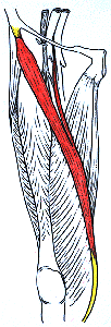
Biceps (Two-Headed) Sartorius
- Laboratory specimen,
University of Iowa. — Anatomy Atlases. Image hosted by the site it comes from. Open the original page.
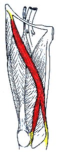
Sartorius Bicaudatus
- Laboratory specimen,
University of Iowa. — Anatomy Atlases. Image hosted by the site it comes from. Open the original page.
Riferimenti dell’opera originale
Elenco riportato come compare nella fonte, senza normalizzazione dei formati bibliografici.
- Bergeron, H. (1866) Muscle couturier double. Bulletins et Mem. de la Société anatomique de Paris XLI(1):2.
- Bhatnagar, B.N.S. and D. Narayan. (1959) Bicipital sartorius. J. Anat. Soc. India 8:32-33.
- Brock, G.S. (1879) A two headed sartorius. J. Anat. Physiol. 13:578.
- Henle, J. (1871) Handbuch der Muskellehre des Menschen, in Handbuch der systematischen Anatomie. Verlag von Friedrich Vieweg un Sohn, Braunschweig.
- Macalister, A. (1871) Observations on muscular anomalies in the human anatomy. Third series with a catalog of muscular variations hitherto published. Trans. Roy. Irish Acad. Sci. 25:1-130.
- Miyasaki, M., Hirohashi, A., Yoshikawa, H. and Y. Nishio. (1958) One case of the variation of the sartorius. Kurume Igakkai Zasshi 21:671-674. In Japanese.
- Mori, M. (1964) Statistics on the musculature of the Japanese. Okajimas Fol. Anat. Jap. 40:195-300.
- Morita, M. (1947) One case of the variation of the M. sartorius. Kaibogaku Zasshi 21:3. In Japanese.
- Nakano, C. (1913) Rare variations of the muscles observed in the dissecting room (M. sartorius and muscles of the arm). Juzenkai Zasshi 18:10-13.
- Schaefer, E.A, Symington, J. and T.H. Bryce., Eds. (1923) Quain's Essentials of Human Anatomy.11th ed. Longmans, Green and Co., London.
Anatomical classification
- Systems:
- Muscular system
Content provenance
Adaptation
Adaptation of «Sartorius — Illustrated Encyclopedia of Human Anatomic Variation: Opus I: Muscular System» by Ronald A. Bergman, Adel K. Afifi, Ryosuke Miyauchi. Source: https://www.anatomyatlases.org/AnatomicVariants/MuscularSystem/Text/S/03Sartorius.shtml Original work distributed under the CC BY-NC-SA 4.0 licence (https://creativecommons.org/licenses/by-nc-sa/4.0/deed.it). Adaptation by Redazione AIVA. Changes from the original: Testo originale inglese riprodotto integralmente e riorganizzato nel formato scheda del portale (sintesi, testo, riferimenti), senza modificarne le affermazioni. La traduzione italiana non è ancora stata eseguita, e la pagina lo dichiara. Le figure non sono state copiate: sono collegate alla pagina d’origine. This publication is not approved or endorsed by the authors of the original work.
Licence
This content is distributed under the licence CC BY-NC-SA 4.0. Licences and attribution