Encyclopedia entry
Supracostalis (Wood)
Abstract
Slips are occasionally found that extend from the ventral end of the first rib to the fourth. Other slips may terminate on the second or third rib. M. supracostalis has also been found attached to the deep fascia of the neck or to the scalene muscles. Supracostalis anterior is found on the anterior surface of the thoracic wall. It usually passes from the anterior end of the first rib downward to the third or fourth rib. Mori reported 126 supracostalis anterior muscles in 1200 cadavers (2400 sides). The form of the muscle is simple but the origin and insertion are variable.
Slips are occasionally found that extend from the ventral end of the first rib to the fourth. Other slips may terminate on the second or third rib. M. supracostalis has also been found attached to the deep fascia of the neck or to the scalene muscles. Supracostalis anterior is found on the anterior surface of the thoracic wall. It usually passes from the anterior end of the first rib downward to the third or fourth rib. Mori reported 126 supracostalis anterior muscles in 1200 cadavers (2400 sides). The form of the muscle is simple but the origin and insertion are variable.
Origin
Highest rib
Number of sides
%
I
105
4.4
II
21
0.8
Insertion
Lowest rib
Number of sides
%
III
58
2.4
IV
68
2.8
Extent of muscle
Lowest rib
Highest Rib
I
II
II
58-2.4%
-
IV
47-1.9%
21-0.8%
According to Mori this muscle has a frequency of 5.2% in 2400 sides. Still other slips, e.g., supracostalis posterior (dorsalis) may be placed dorsal to the origin of serratus anterior. They may occur singly or in multiples, bilaterally or unilaterally. They are classed along with the serratus posterior muscles as remnants of the superficial lamellae of the primitive external oblique muscle. Supracostalis posterior is considered to occur in three types according to Eisler (cited by Mori): 1). Located on the dorsal wall of the thorax, its component muscle fibers follow the same direction as the external intercostal muscle. It arises and inserts on ribs, its length determined by whether one or two ribs are skipped. 2). The muscle fibers may arise from ribs as one to three fleshy slips or digitations. The muscle fibers have the same direction as serratus posterior superior (craniomedial direction). 3). The muscle bundles arise from the eighth to tenth ribs and pass in a craniomedial direction. The insertion depends on the degree of development of the muscle bundles. When poorly developed the muscle fibers at the cranial end of the muscle join the fascia over the ribs. When highly developed, the terminal tendon inserts into the fascia of the deep muscles of the trunk (erector spinae). Mori reported 65 cases of supracostalis dorsalis as follows: Type 1... 19 sides... 3.6%. Type 2 ... 17 sides ... 3.3%. Type 3 ... 29 sides ... 5.6%.
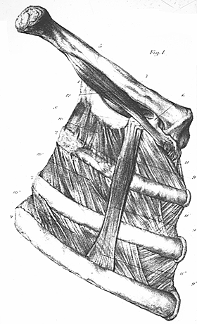
Supracostalis Anterior
from Bochdalek, Jr., 1867 — Anatomy Atlases. Image hosted by the site it comes from. Open the original page.

Supracostalis Posterior
From Mori, 1964 — Anatomy Atlases. Image hosted by the site it comes from. Open the original page.
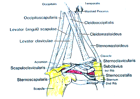
Varieties of Chest, Neck, and Shoulder Muscles-Sternoscapularis.
- modified and redrawn from Wood, 1870.
Anatomy Atlases. Image hosted by the site it comes from. Open the original page.
Riferimenti dell’opera originale
Elenco riportato come compare nella fonte, senza normalizzazione dei formati bibliografici.
- Bochdalek, Jr., -. (1867) Ein anomaler Musculus supracostalis anterior. Arch. Path. Anat. Physiol. Klin. Med. 41:257-258.
- Keith, A.M. (1894) Note on supracostalis anterior. J. Anat. Physiol. 27:333-334.
- Kurotsu, T. (1932) On the M. supracostalis posterior. Kaibogaku Zasshi 5: 4 42-447. In Japanese.
- Livini, F. (1905) Contribuzione alla morfologia des m. rectus abdominis e del m. supra-costalis nell'uomo. Arch. Ital. Anat. Embriol. 4:81-116.
- Livini, F. (1905) Contribution à la morphologie du m. rectus abdominis et du m. supracostalus chexz l'homme. Arch. Ital. Biol. 44:116-117.
- Macalister, A. (1875) Observations on the muscular anomalies in the human anatomy. Third series with a catalogue of the principal muscular variations hitherto published. Trans. Roy. Irish Acad. Sci. 25:1-130.
- Miyauchi, R. (1976) Four cases of rare supernumerary muscles that were found on the anterior chest wall. Medical Bulletin of Fukuoka University. 3:353-365.
- Miyauchi, R. (1986) A case of a rare anomalous muscle of the human anterior thoracic wall. Okajimas Folia Anatomica Japonica 63:331-336.
- Mori, M. (1964) Statistics on the musculature of the Japanese. Okajimas Folia Anatomica Japonica 40:195-300.
- Mori, M. and M. Nazayoshi. (1938) Two cases of the M. supracostalis anterior. Kyushi Gakuho 2:8-9. In Japanese.
- Pye-Smith, -. (1868) Ein zweiter Fall von Musculus supracostalis anterior anomalous. Arch. Path. Anat. Physiol. Klin. Med. 43:142.
- Wood, J. (1865) Additional varieties in human myology. Proc. Roy. Soc. Lond. B 14:378-392.
- Wood, J. (1870) On a group of varieties of the muscles of the human neck, shoulder, and chest, and their transitional forms and homologies in the mammalia. Phil. Trans. R. Soc. (Lond.) 160:83-116.
Anatomical classification
- Systems:
- Muscular system
Content provenance
Adaptation
Adaptation of «Supracostalis (Wood) — Illustrated Encyclopedia of Human Anatomic Variation: Opus I: Muscular System» by Ronald A. Bergman, Adel K. Afifi, Ryosuke Miyauchi. Source: https://www.anatomyatlases.org/AnatomicVariants/MuscularSystem/Text/S/57Supracostalis.shtml Original work distributed under the CC BY-NC-SA 4.0 licence (https://creativecommons.org/licenses/by-nc-sa/4.0/deed.it). Adaptation by Redazione AIVA. Changes from the original: Testo originale inglese riprodotto integralmente e riorganizzato nel formato scheda del portale (sintesi, testo, riferimenti), senza modificarne le affermazioni. La traduzione italiana non è ancora stata eseguita, e la pagina lo dichiara. Le figure non sono state copiate: sono collegate alla pagina d’origine. This publication is not approved or endorsed by the authors of the original work.
Licence
This content is distributed under the licence CC BY-NC-SA 4.0. Licences and attribution