Encyclopedia entry
Tibialis Posterior
Abstract
Variations of this muscle are few and rare. At least two cases have been reported in which the muscle consisted of two parts; the accessory part was inserted separately in the sole of the foot. The insertion onto the first cuneiform may be absent. Macalister reported the variations of the tibialis posterior as follows: 1. It may have a trifurcated tendon, one slip going to the usual site of insertion, the second to the tendon of peroneus longus, and the third to the heads of the third and fourth metatarsal bones; 2.
Variations of this muscle are few and rare. At least two cases have been reported in which the muscle consisted of two parts; the accessory part was inserted separately in the sole of the foot. The insertion onto the first cuneiform may be absent.
Macalister reported the variations of the tibialis posterior as follows: 1. It may have a trifurcated tendon, one slip going to the usual site of insertion, the second to the tendon of peroneus longus, and the third to the heads of the third and fourth metatarsal bones; 2. The slip that frequently goes to the ento-cuneiform (intermediate cuneiform) may give attachment to the flexor hallucis by joining it; 3. It may supply a tendinous slip to the second and third metatarsal (Harrison); 4. Its absence has been reported by Budge.
Another muscle, tibialis secundus, arises from the posterior aspect of the tibia below tibialis posterior and inserts into the capsule of the ankel joint. A similar slip (tensor capsuli tibiotarsalis anterior) , ending on the anterior wall of the capsule of the ankel joint, is considered by some investigators to be a variant of the long accessory muscle described.
Tibiotarsalis arises from the popliteal line and back of the tibia and inserts into the plantar fascia. This muscle is considered to be a tensor fasciae plantaris.
The tendon of tibialis posterior may send a slip to the tendon of flexor hallucis brevis, which may give rise to hallux valgus.
Syn.: m. tibiaeus posticus, tibialis posterior, tibiaeus nauticus, Schwimmuskel (Theile).
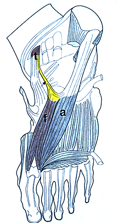
Expansion of Tibialis Posterior tendon to Oblique Part of Adductor Hallucis Muscle.
- a, oblique part of adductor hallucis; f, flexor hallucis brevis; t, tibialis posterior tendon. Arrow, deviant tendinous expansion; arrow head, deviated great toe.
from Gunal, Sahinoglu, and Bergman. — Anatomy Atlases. Image hosted by the site it comes from. Open the original page.
Riferimenti dell’opera originale
Elenco riportato come compare nella fonte, senza normalizzazione dei formati bibliografici.
- Anderson, R.J. (1897) Vorkommen eines Musculus tibio-tarsalis sive tensor fasciae plantaris. Arch. Path. Anat. Physiol. Klin. Med. 81:574-575.
- Anson, B.J., Ed. (1966) Morris' Human Anatomy,12th ed. The Blakiston Division, McGraw-Hill Book Company, New York.
- Gunal, I., Sahinoglu, K. and R.A. Bergman. (1994) Anomalous tibialis posterior muscle as an etiologic factor of hallux valgus. Clinical Anat. 7(1):21-25.
- Macalister, A. (1875) Observations on the muscular variations in the human anatomy. Third series with a catalogue of the principal muscular variations hitherto published. Trans. Roy. Irish Acad. Sci. 25:1-130.
- Musial, W.W. (1963) Variations of the terminal insertions of the anterior and posterior tibial muscles in man. Folia Morphol. (Warsaw) 22:294-302.
- Toldt, C. (1928) An atlas of Human Anatomy for Students and Physicians. The Macmillian Company, New York.
Anatomical classification
- Systems:
- Muscular system
Content provenance
Adaptation
Adaptation of «Tibialis Posterior — Illustrated Encyclopedia of Human Anatomic Variation: Opus I: Muscular System» by Ronald A. Bergman, Adel K. Afifi, Ryosuke Miyauchi. Source: https://www.anatomyatlases.org/AnatomicVariants/MuscularSystem/Text/T/24Tibialis.shtml Original work distributed under the CC BY-NC-SA 4.0 licence (https://creativecommons.org/licenses/by-nc-sa/4.0/deed.it). Adaptation by Redazione AIVA. Changes from the original: Testo originale inglese riprodotto integralmente e riorganizzato nel formato scheda del portale (sintesi, testo, riferimenti), senza modificarne le affermazioni. La traduzione italiana non è ancora stata eseguita, e la pagina lo dichiara. Le figure non sono state copiate: sono collegate alla pagina d’origine. This publication is not approved or endorsed by the authors of the original work.
Licence
This content is distributed under the licence CC BY-NC-SA 4.0. Licences and attribution