Encyclopedia entry
Transversus Thoracis Anterior (Sternocostalis) and Subcostalis
Abstract
Transversus thoracis arises from the posterior surface of the sternum and xiphoid (to the level of the third costal cartilage) and extends onto the internal surface of the second (third) to sixth costal cartilages. Transversus thoracis may extend to the first rib. Absence on one or both sides has been recorded. It is sometimes divided into separate fasciculi which may number from two to six, and its division into two lamellae has also been observed. This muscle was Hyrtl's candidate for the most variable in the body.
Transversus thoracis arises from the posterior surface of the sternum and xiphoid (to the level of the third costal cartilage) and extends onto the internal surface of the second (third) to sixth costal cartilages. Transversus thoracis may extend to the first rib. Absence on one or both sides has been recorded. It is sometimes divided into separate fasciculi which may number from two to six, and its division into two lamellae has also been observed. This muscle was Hyrtl's candidate for the most variable in the body.
Mori provides a statistical report of 310 sides, 155 cadavers:
Highest Rib of Insertion
Rib
Number of sides
%
I
7
2.2
II
122
39.3
III
155
50.0
IV
23
7.4
V
3
0.9
Lowest Rib of Insertion
Rib
Number of sides
%
V
50
16.1
VI
237
76.4
VII
23
7.4
The Extent of Transversus Thoracis on Inner Surface of Thoracic Wall
Highest Rib
Lowest Rib
V
VI
VII
I
1 (0.3%)
6 (3.9%)
9 (2.9%)
II
27 (8.74%)
86 (27.7%)
-
III
21 (6.7%)
121 (39.3%)
13 (4.1%)
IV
1 (.3%)
21 (6.7%)
1 (0.3%)
V
3 (0.9%)
The subcostales are usually better developed on the inner surface of the thorax and are variable in number.
Syn.: Tr. Th.: m. triangularis sterni s. sternocostalis (Henle), transversus pectoris (Arnold), Sternoabdominalis (Rosenmüller), Dreieckiger or innerer Brustmuskel, Petit dentelé anterieur (Cruveilheir).
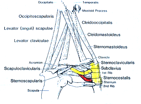
Varieties of Chest, Neck, and Shoulder Muscles-Sternocostalis.
- modified and redrawn from Wood.
Anatomy Atlases. Image hosted by the site it comes from. Open the original page.
Riferimenti dell’opera originale
Elenco riportato come compare nella fonte, senza normalizzazione dei formati bibliografici.
- Anson, B.J., Ed. (1966) Morris' Human Anatomy, 12th ed. The Blakiston Division, McGraw-Hill Book Company, New York.
- Macalister, A. (1875) Observations on the muscular anomalies in the human anatomy. Third series with a catalogue of the principal muscular variations hitherto published. Trans. Roy. Irish Acad. Sci. 25:1-134.
- Mori,M. (1964) Statistics on the musculature of the Japanese. Okajimas Fol. Anat. Jap. 40:195-300.
- de Pina, L. (1933) Le muscle petit dentele postieur et superieur chez l'homme et les primates. L'Assoc. Anatomistes, Comptes rendus 28:523-531.
- Wood, J. (1870) On a group of varieties of the muscles of the human neck, shoulder, and chest, and their transitional forms and homologies in the mammalia. Phil. Trans. R. Soc. (Lond.) 160:83-116.
Anatomical classification
- Systems:
- Muscular system
Content provenance
Adaptation
Adaptation of «Transversus Thoracis Anterior (Sternocostalis) and Subcostalis — Illustrated Encyclopedia of Human Anatomic Variation: Opus I: Muscular System» by Ronald A. Bergman, Adel K. Afifi, Ryosuke Miyauchi. Source: https://www.anatomyatlases.org/AnatomicVariants/MuscularSystem/Text/T/45Transversus.shtml Original work distributed under the CC BY-NC-SA 4.0 licence (https://creativecommons.org/licenses/by-nc-sa/4.0/deed.it). Adaptation by Redazione AIVA. Changes from the original: Testo originale inglese riprodotto integralmente e riorganizzato nel formato scheda del portale (sintesi, testo, riferimenti), senza modificarne le affermazioni. La traduzione italiana non è ancora stata eseguita, e la pagina lo dichiara. Le figure non sono state copiate: sono collegate alla pagina d’origine. This publication is not approved or endorsed by the authors of the original work.
Licence
This content is distributed under the licence CC BY-NC-SA 4.0. Licences and attribution