Encyclopedia entry
Vagus (CN X) Nerve
Abstract
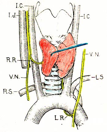
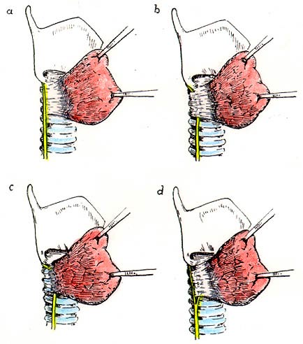
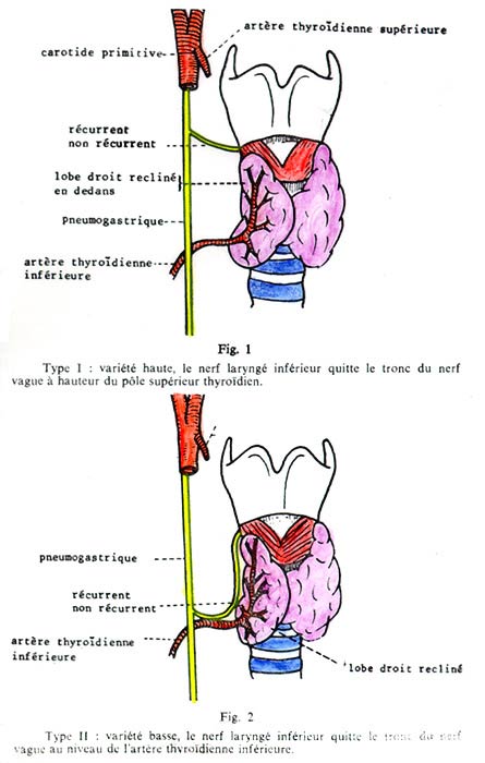
The right and left vagus may differ significantly in size. The nerves may lie in front of both vessels (internal carotid and internal jugular) in the carotid sheath. The left vagus has been found passing anterior to the common carotid artery and the thyroid gland. Descendens hypoglossi (C1 and C2) may be carried partly or completely by the vagus nerve instead of the hypoglossal nerve. The distribution of the right and left vagus nerves around the esophageal hiatus is variable. The vagus nerve may contain accessory parathyroid tissue. See also Recurrent Laryngeal Nerve.
The right and left vagus may differ significantly in size.
The nerves may lie in front of both vessels (internal carotid and internal jugular) in the carotid sheath.
The left vagus has been found passing anterior to the common carotid artery and the thyroid gland.
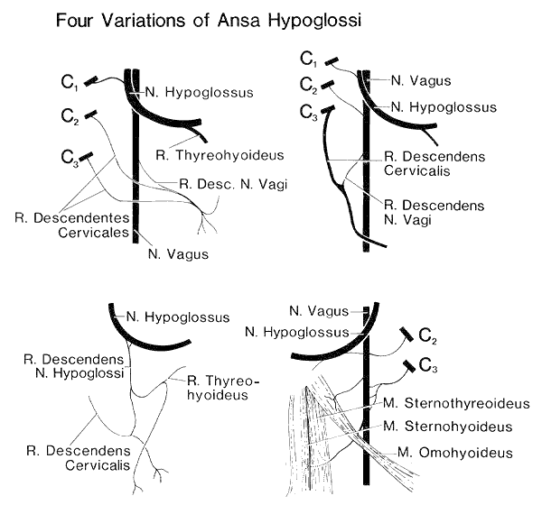
Descendens hypoglossi (C1 and C2) may be carried partly or completely by the vagus nerve instead of the hypoglossal nerve.
The distribution of the right and left vagus nerves around the esophageal hiatus is variable.
The vagus nerve may contain accessory parathyroid tissue.
See also Recurrent Laryngeal Nerve.
Image 27A Image 27B Image 27C Image 27D
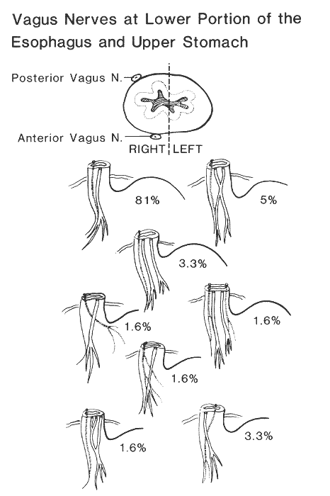
Anatomy Atlases. Image hosted by the site it comes from. Open the original page.
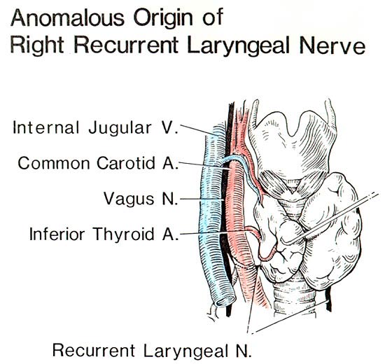
Anatomy Atlases. Image hosted by the site it comes from. Open the original page.
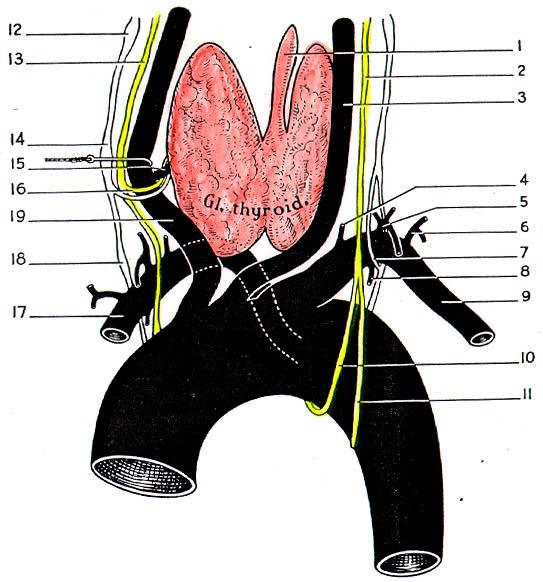
Anatomy Atlases. Image hosted by the site it comes from. Open the original page.
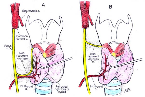
Anatomy Atlases. Image hosted by the site it comes from. Open the original page.
Anatomy Atlases. Image hosted by the site it comes from. Open the original page.
Image 27E Image 27F Image 73A Image 73B
Anatomy Atlases. Image hosted by the site it comes from. Open the original page.
Anatomy Atlases. Image hosted by the site it comes from. Open the original page.
Anatomy Atlases. Image hosted by the site it comes from. Open the original page.
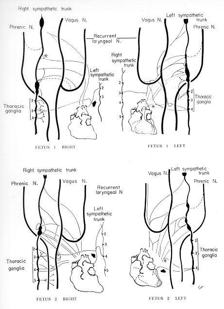
Anatomy Atlases. Image hosted by the site it comes from. Open the original page.
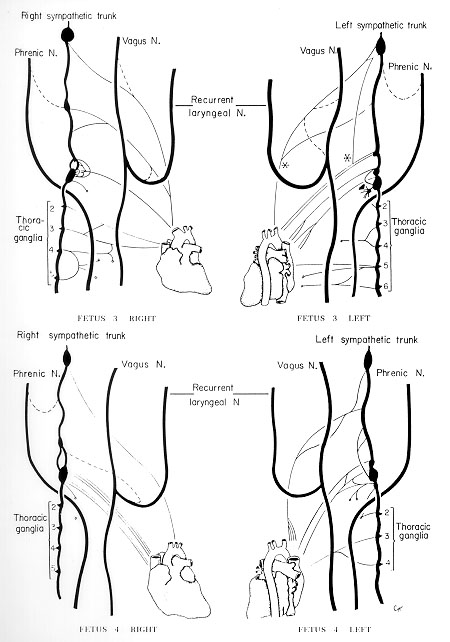
Anatomy Atlases. Image hosted by the site it comes from. Open the original page.
Image 73C Image 73D Image 73E Image 73F
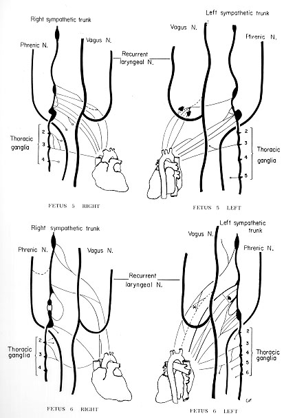
Anatomy Atlases. Image hosted by the site it comes from. Open the original page.

Anatomy Atlases. Image hosted by the site it comes from. Open the original page.
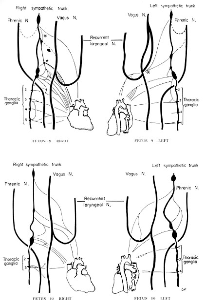
Anatomy Atlases. Image hosted by the site it comes from. Open the original page.
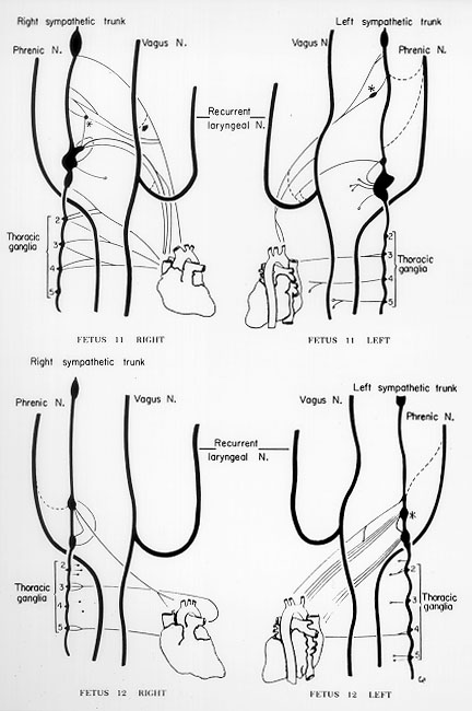
Anatomy Atlases. Image hosted by the site it comes from. Open the original page.
Riferimenti dell’opera originale
Elenco riportato come compare nella fonte, senza normalizzazione dei formati bibliografici.
- Anson, B.J., Ed. (1966) Morris' Human Anatomy, 12th ed., The Blakiston Division, McGraw-Hill Book Company, New York.
- Chamberlin, J.A. and T Winship. (1947) Anatomic variations of the vagus nerves - their significance in vagus neurectomy. Surgery 22:1-19.
- Cruveilhier, J. (1851) Traité d'Anatomie Descriptive, 3rd ed., G. Doin & Cie., Paris.
- Doubilet, H., Shafiroff, B.G.P. and J.H. Mulholland. (1948) The anatomy of the periesophageal vagi. Ann. Surgery 127:128-135.
- Dragstedt, L.R., Fournier, H.J., Woodward, E.R., Tovee, E.B. and P.V. Harper, Jr. (1947). Transabdominal gastric vagotomy. study of the anatomy and surgery of the vagus nerves at the lower portion of the esophagus. Surg. Gynecol. Obstet. 85:461-466.
- Gibson, A. (1915) Bilateral abnormal relationship of the vagus nerve in its cervical portion. J. Anat. Physiol. 69:389-392.
- Henle, J. (1868) Handbuch der Systematischen Anatomie des Menschen., von Friedrich Vieweg und Sohn, Braunschweig.
- Huu, N. (1954) Les nerfs de Telford, Stopford et Mitchell et la problème de la terminasion du pneumogastrique. Première note: Les arguments anatomiques. Assoc. Anatomistes Comptes Rendus 41:821-830.
- Jo, A. (1971) Eine seltene Anastomose des Nervus vagus. Anat. Anz. 129:180-182.
- Kopsch, F. (1908) Rauber's Lehrbuch der Anatomie des Menschen., Georg Thieme, Leipzig.
- Kurylcio, L. (1971) Forms of the vagus trunk and their distribution in the esophageal hiatus in man. Folia Morphol., Warsaw 30:73-85.
- Kurylcio, L. (1972) Anatomic varieties of the vagus trunks from the point of view of selective vagotomy. Folia Morphol., (Warsaw) 31:85-108.
- Kurylcio, L. (1972) The terminal branches of the vagus trunks in man. Folia Morphol., (Warsaw) 31:231-259.
- Latarjet, A. (1948) Testut's Traité d'Anatomie Humaine., 9th ed., G. Doin & Cie., Paris.
- Lippmann, R. (1910) Ursprung des Ramus descendens n. hypoglossi aus dem N. vagi. Anat. Anz. 37:1-4.
- Mirto, D. e E. Pulsateri. (1896) Sue les rapports anastomotiques entre le nerf accessoire et le vague. Arch. Ital. Biol. 25:348.
- Romanes, G.J. (1953) The distribution of taste buds on the human epiglottis. J. Anat. 87:464.
- Schaefer, E.A., Symington, J. and T.H. Bryce., Eds. (1915) Quain's Anatomy, 11th ed., Longmans, Green, and Co., London.
- Taguchi, K. (1888) über eine seltene Anaomalie des Verlaufes des Vagusstammes und eine seiner Äste. Arch. Anat. Physiol. Wissen. Med. 1888:365-368.
- Willm, A. (1928) Absence totale de l'anse de l'hypoglosse, les nerfs des muscles soushyoïdiens naissant en apparence de pneumogastrique. Ann. Anat. Pathol. 5:580-582.
Anatomical classification
- Systems:
- Nervous system
Content provenance
Adaptation
Adaptation of «Vagus (CN X) Nerve — Illustrated Encyclopedia of Human Anatomic Variation: Opus I: Muscular System» by Ronald A. Bergman, Adel K. Afifi, Ryosuke Miyauchi. Source: https://www.anatomyatlases.org/AnatomicVariants/NervousSystem/Text/VagusNerve.shtml Original work distributed under the CC BY-NC-SA 4.0 licence (https://creativecommons.org/licenses/by-nc-sa/4.0/deed.it). Adaptation by Redazione AIVA. Changes from the original: Testo originale inglese riprodotto integralmente e riorganizzato nel formato scheda del portale (sintesi, testo, riferimenti), senza modificarne le affermazioni. La traduzione italiana non è ancora stata eseguita, e la pagina lo dichiara. Le figure non sono state copiate: sono collegate alla pagina d’origine. This publication is not approved or endorsed by the authors of the original work.
Licence
This content is distributed under the licence CC BY-NC-SA 4.0. Licences and attribution