Encyclopedia entry
Variations in Size and in Symmetry of Foramina of the Human Skull
Abstract
Table 1 Paired Foramina Foramen Average Size (mm) Largest Size (mm) Smallst Size (mm) Anterior Ethmoid 2.27 x 1.14 8 x 3 0.64 x 0.64 Carotid Canal 7.13 x 5.29 12 x 5 5.5 x 4 Foramen Ovale 7.11 x 3.60 8 x 7 5 x 2 Foramen Rotundum 3.26 x 2.66 4.5 x 3.5 2 x 2 Foramen Spinosum 2.39 x 1.96 4 x 3 1 x 1 Greater Palatine 4.01 x 2.16 8 x 3 1.67 x 1.67 Hiatus for the Greater Petrosal 0.77 x 0.68 3 x 2 0.3 x 0.3 Hiatus for the Lesser Petrosal 0.37 x 0.37 0.83 x 0.83 0.3 x 0.3 Hypoglossal (Exocranial) 4.66 x 3.21 7.5 x 5 2 x 0.92 Infraorbital Foramen 3.42 x 2.40 6 x 4 1.67 x 1.67 Innominate 2.13 x 1.1 4 x…
Table 1 Paired Foramina
Foramen
Average Size (mm)
Largest Size (mm)
Smallst Size (mm)
Anterior Ethmoid 2.27 x 1.14 8 x 3 0.64 x 0.64
Carotid Canal 7.13 x 5.29 12 x 5 5.5 x 4
Foramen Ovale 7.11 x 3.60 8 x 7 5 x 2
Foramen Rotundum 3.26 x 2.66 4.5 x 3.5 2 x 2
Foramen Spinosum 2.39 x 1.96 4 x 3 1 x 1
Greater Palatine 4.01 x 2.16 8 x 3 1.67 x 1.67
Hiatus for the Greater Petrosal 0.77 x 0.68 3 x 2 0.3 x 0.3
Hiatus for the Lesser Petrosal 0.37 x 0.37 0.83 x 0.83 0.3 x 0.3
Hypoglossal (Exocranial) 4.66 x 3.21 7.5 x 5 2 x 0.92
Infraorbital Foramen 3.42 x 2.40 6 x 4 1.67 x 1.67
Innominate 2.13 x 1.1 4 x 3 0.3 x 0.3
Internal Acoustic Meatus 4.27 x 3.78 6 x 5 3 x 3
Lacrimal 0.71 x 0.67 4 x 1 0.3 x 0.3
Lesser Palatine 0.86 x 0.73 3.5 x 1.5 0.3 x 0.3
Mandibular 3.29 x 3.51 6.5 x 6 1 x 1
Mastoid Foramen 1.26 x 1.09 5.5 x 5 0.3 x 0.3
Mental Foramen 2.43 x 1.76 5.5 x 3 0.52 x 0.52
Nasolacrimal 5.30 x 4.45 7 x 7 3.5 x 3.5
Optic Foramen 5.02 x 4.16 7 x 5 3.5 x 3
Parietal foramina permagna 0.67 x 0.67 1.67 x 1.67 0.3 x 0.3
Post. Condylar 2.94 x 2.56 6 x 4.5 0.41 x 0.41
Posterior Ethmoid 1.23 x 0.82 4 x 1.5 0.41 x 0.41
Stylomastoid 1.66 x 1.56 2.5 x 2 1.07 x 1.07
Superior Alveolar 0.57 x 0.57 1.07 x 1.07 0.3 x 0.3
Superior Orbital 1.85 x 1.15 3.5 x 1.5 0.52 x 0.52
Trochlear 0.76 x 0.73 1.28 x 0.83 0.3 x 0.3
Vesalius 0.79 x 0.59 3 x 2 0.3 x 0.3
Unpaired Foramina
Foramen Magnum 33.8 x 28.3 41 x 53 24 x 23
Incisive Canal Length 2.76 x 2.39 5.5 x 4 0.46 x 0.46
Canal Average Length (mm) Longest Length (mm) Shortest Length (mm)
Infraorbital Canal 27.7 35 22
There was no significant difference between the average size of the foramina on the right and the average size of the foramina on the left, for any pair of foramina. Therefore, the "average size" was calculated by taking the average of both the right and left for each pair of foramina. When multiple openings were present for a foramen, all the openings were measured and used in the calculation of the average size. Table 2 Symmetry of Paired Foramina
Foramen
Absent Bilateral
Present Unilateral
Bilateral Same Size
Bilateral >0.5mm Diff
Anterior Ethmoid 0/90(0%) 1/90(1%) 65/90(72%) 24/90(27%)
Carotid Canal 0/100(0%) 0/100(0%) 77/100/(77%) 23/100(23%)
Foramen Ovale 0/96(0%) 0/96(0%) 40/96(42%) 56/96(58%)
Foramen Rotundum 0/98(0%) 0/98(0%) 90/98(92%) 8/98(8%)
Foramen Spinosum 0/92(0%) 1/99(1%) 77/92(84%) 14/92(15%)
Greater Palatine 0/99(0%) 0/99(0%) 67/99(68%) 32/99(32%)
Hiatus for the Greater Petrosal 0/100(0%) 0/100(0%) 87/100(87%) 13/100(13%)
Hiatus for the Lesser Petrosal 15/100(15%) 11/100(11%) 72/100(72%) 2/100(2%)
Hypoglassal (Exocranial) 0/100(0%) 0/100(0%) 41/100(41%) 59/100(100%)
Infraorbital 0/98(0%) 0/98(0%) 73/98(74%) 25/98(26%)
Innominate 65/100(65%) 17/100(17%) 9/100(9%) 9/100(9%)
Internal Acoustic Meatus 0/100(0%) 0/100(0%) 91/100(91%) 9/100(9%)
Lacrimal 28/100(28%) 31/100(31%) 35/100(35%) 6/100(6%)
Lesser Palatine 0/99(0%) 3/99(3%) 77/99(78%) 19/99(19%)
Mandibular 0/100(0%) 0/100(0%) 92/100(92%) 8/100(8%)
Mastoid 1/99(1%) 10/99(10%) 47/99(47%) 41/99(41%)
Mental 0/100(0%) 0/100(0%) 80/100(80%) 20/100(20%)
Nasolacrimal 0/46(0%) 0/46(0%) 38/46(83%) 8/46(17%)
Optic Canal 0/97(0%) 0/97(0%) 82/97(85%) 15/97(15%)
Parietal 20/99(20%) 30/99(30%) 33/99(33%) 16/99(16%)
Post. Condylar 10/100(10%) 36/100(36%) 18/100(18%) 36/100(36%)
Posterior Ethmoid 0/98(0%) 0/98(0%) 78/98(80%) 20/98(20%)
Stylomastoid 0/99(0%) 0/99(0%) 98/99(99%) 1/99(1%)
Superior Alveolar 1/91(1%) 0/91(0%) 86/91(95%) 4/91(4%)
Superior Orbital 50/100(50%) 31/100(31%) 14/100(14%) 5/100(5%)
Trochlear 92/100(92%) 6/100(6%) 2/100(2%) 0/100(0%)
Vesalius 41/100(41%) 24/100(24%) 25/100(25%) 10/100(10%)
Infraorbital Canal 0/98(0%) 0/98(0%) 64/98(65%) 34/98(35%)
Although 100 skulls were examined, not every foramen in the skull was useable due to breakage or inaccessibility. The number of useable skulls for each pair of foramina is given as the denominator. The four colums represent:
- the number of skulls in which neither foramina of the pair was present
- the number of skulls in which only one of the two foramina of the pair was present
- the number of skulls in which both of the foramina of the pair were present and the same size within 0.5 mm
- the number of skulls in which both of the foramina were present but there was greater than 0.5 mm difference in the size of the two foramina.
Variation in the Shape of the Superior Orbital Fissure
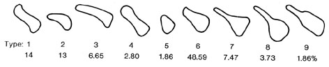
Anatomy Atlases. Image hosted by the site it comes from. Open the original page.
Riferimenti dell’opera originale
Elenco riportato come compare nella fonte, senza normalizzazione dei formati bibliografici.
- Bergman R.A., Thompson S.A., Afifi A.K., Saadeh F.A. (1988) Compendium of Human Anatomic Variation; World Literature, Catalog and Atlas. Urban and Schwarzenberg, Munich and Baltimore.
- Berlis, A., Putz, R. and M. Schumacher. (1992) Direct and CT measurements of canals and foramina of the skull base. The British Journal of Radiology 65:653-661.
- Berry A.C., Berry R.J. (1967) Epigenetic variation in the human cranium. J. Anat. 101:361-379.
- Berry A.C. (1975) Factors affecting the incidence of non-metrical skeletal variants. I Anat. 120, 3:519-535.
- Boyd G.I. (1930) Emilssary foramina of cranium in man and anthropoids. J. Anat. 65:108-121.
- Coin C.G., Malkasian D.R. (197 1) Foramen magnum. In: Newton T.H., Potts D.G., eds. Radiology of the skull and brain. St. Louis: Mosby, 1:1 275-286.
- Currarino G,, Maravilla K.R., Salver K.E. (1985) Transsphenoidal canal and its pathologic implications, ANJR 6:39-43.
- Curtin A.D., Williams R., Johnson J. (1984) CT of perineural tumor extension: pterygopalatine fossa, AJNR 5:731-737.
- Goalwain H.A. (1927ab.) One thousand optic canals, J. Am. Med. Assoc. 89:1745. -- (1927b) The profile roentgenogram of the optic canal, Am. J. Roentgenol. 17:573.
- Henderson W.R. (1966) A note on the relationship of the human maxillary nerve to the cavernous sinus and to an emissary sinus passing through the foramen ovale. J. Anat. 100:905-908.
- Lanzieri C.F., Duchesneau P.M., Rosenbloom S.A., Smith A.S., Rosenbaum A.E. (1988) The signficance of asymmetry of the foramen of Vesalius. AJNR 9:1201-1204.
- Le Double A.F. (1903) Traité des variations des os du crâne de l'homme er de leur signification au pont de vue de l'anthroppologie zoologique, Paris, Vigot Frères.
- Lindblom K. (1936) A roentgenographic study of the vascular channels of the skull, Acta Radiol. supp 30.
- Pandolfa, I., Gaeta M., Blandino A., Longo M. (1987) The radiology of the pterygoid canal: normal and pathological findings, AJNR 8:479-483.
- Radoïévitch A., Jovanovich A., Lotritch N. (1956) La morphologie dur trou ovale et les rapports du nerf maxilaire inférieur avec le sinus sphénoidal, Rev. Laryng. 77:11-19.
- Russell F. (1900) Studies in cranial variations, Amer. Nat. 34:737-747.
- Shapiro R., Robinson F. (1967) The foramina of the middle fossa: a phylogenetic, anatomic and, pathologic study, Amer. J. Roentgen. 101:779-794.
- Sondheimer F.K. (1971) Basal foramina and canals. In, Newton T.H., Potts D.G., eds. Radiology of the skull and brain. St..Louis: Mosby 1:1 287-347.
- Sperino, -. (1884) Circolazione Venosa del Dapo. Turin (cited by Boyd 1903).
- Streit H. (1903) Über otologisch wichtige Anomalien der Hirnsinus, über acessorische sinus und bedeutendere Venenverbindungen. Arch. Ohrenheilk. 58:85-128 and 161-167.
- Wang T., Kuo K., Shih C., Ho L., Liu J. (1988) Assessment of the relative locations of the greater palatine foramen in adult Chinese skulls, Acta Anat. 132:182-186.
Anatomical classification
- Systems:
- Skeletal system
Content provenance
Adaptation
Adaptation of «Variations in Size and in Symmetry of Foramina of the Human Skull — Illustrated Encyclopedia of Human Anatomic Variation: Opus I: Muscular System» by Ronald A. Bergman, Adel K. Afifi, Ryosuke Miyauchi. Source: https://www.anatomyatlases.org/AnatomicVariants/SkeletalSystem/Text/VariationsSizeSymmetry.shtml Original work distributed under the CC BY-NC-SA 4.0 licence (https://creativecommons.org/licenses/by-nc-sa/4.0/deed.it). Adaptation by Redazione AIVA. Changes from the original: Testo originale inglese riprodotto integralmente e riorganizzato nel formato scheda del portale (sintesi, testo, riferimenti), senza modificarne le affermazioni. La traduzione italiana non è ancora stata eseguita, e la pagina lo dichiara. Le figure non sono state copiate: sono collegate alla pagina d’origine. This publication is not approved or endorsed by the authors of the original work.
Licence
This content is distributed under the licence CC BY-NC-SA 4.0. Licences and attribution
