Voce enciclopedica
Braccio: omero
Abstract
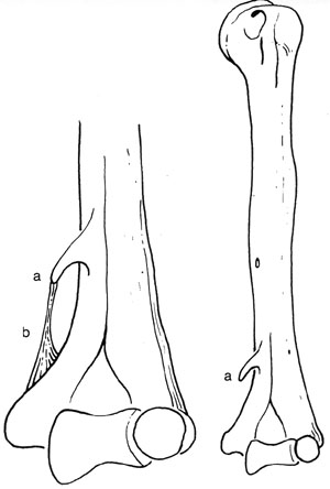
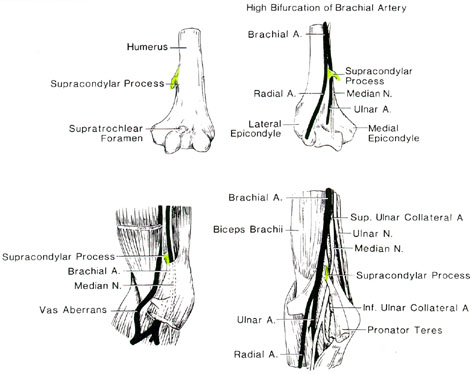
L’omero può presentare una fossa olecranica perforata (forame sovratrocleare) e, sulla superficie anteromediale distale, un processo sopracondiloideo unito all’epicondilo mediale da una banda fibrosa (cosiddetto legamento di Struthers). Il canale così formato trasmette il nervo mediano e l’arteria brachiale, che possono esservi compressi.
La testa dell’omero può essere assente.
L’omero può presentare un processo deltoideo ingrandito.
La fossa olecranica dell’omero è occasionalmente perforata così da formare un forame sovratrocleare, o apertura settale. Le aperture settali possono comparire nel 4-13% degli individui, più frequentemente sul lato sinistro e nelle donne. Sono state riportate variazioni razziali del 4,1-58%. Nell’esame di 1744 ossa del braccio, Trotter riscontrò aperture settali nel 4,2% dei bianchi e nel 12,8% dei neri americani.
Una spina ossea uncinata di dimensioni variabili, il processo sopracondiloideo, può sporgere distalmente dalla superficie anteromediale dell’omero, circa 5 cm prossimalmente all’epicondilo mediale. Esso è unito all’epicondilo da una banda fibrosa (cosiddetto legamento di Struthers) che può ossificarsi. Il processo, la banda e il corpo dell’omero formano un anello o canale attraverso il quale passano il nervo mediano e l’arteria brachiale (o un suo ramo). Il nervo e/o l’arteria possono venire compressi causando sintomi clinici.
Occasionalmente solo il nervo mediano attraversa il canale.
Il processo osseo e il legamento talvolta danno origine a una parte del muscolo pronatore rotondo, oltre a offrire sede di inserzione a una porzione del coracobrachiale.
Il processo sopracondiloideo dell’omero ha una frequenza riportata del 2,7% (Gruber). Adachi riassunse la letteratura (fino al 1828) e trovò che 12 autori (Gruber escluso) avevano studiato 9620 omeri e identificato processi sopracondiloidei in 78, ossia nello 0,8% delle braccia. L’intervallo di frequenza nei 12 studi era 0,1-5,7%. Le popolazioni studiate comprendevano Aino, cinesi, coreani, neri, melanesiani, australiani, giapponesi (inclusi 142 reperti dell’età della pietra), indiani orientali, tedeschi, francesi, eschimesi, svedesi e italiani.
Gruber (1865) osservò: «questa conoscenza non può essere del tutto priva di interesse per il chirurgo».
Un osso sesamoide si riscontra frequentemente nel tendine di inserzione del muscolo tricipite.
Robert Spinner e coll. rilevano che Tiedemann descrisse per primo il processo nel 1818, seguito da Knox nel 1841 e da Struthers nel 1849.
Processo sopracondiloideo
Anatomy Atlases. Immagine ospitata dal sito d’origine. Apri la pagina originale.
Processo sopracondiloideo. Forame sovratrocleare.
Anatomy Atlases. Immagine ospitata dal sito d’origine. Apri la pagina originale.
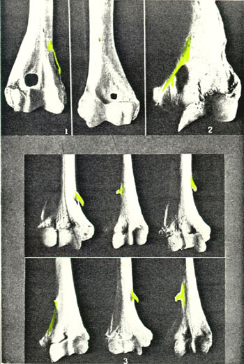
Vedute dell’estremità superiore dell’omero sinistro
Anatomy Atlases. Immagine ospitata dal sito d’origine. Apri la pagina originale.
I complessi strutturali di Struthers del braccio
Riferimenti dell’opera originale
Elenco riportato come compare nella fonte, senza normalizzazione dei formati bibliografici. Le voci non sono tradotte: un riferimento bibliografico si cita nella lingua in cui è pubblicato.
- Akabori, E. (1934) Septal aperatures in the humerus in Japanese, Ainu, and Koreans. Am. J. Phys. Anthropol. 18:395-500.
- Akabori, E. (1935) On the aperture in the fossa olecrani of some east Asiatics. Folia Anat. Jpn. 13:473-475.
- Albers-Schönberg, -. (1915-16) Eine seltene, bisher nicht bekannte Strukturanomalie des Skelettes. Skelettanomalie von atavistischem Interesse, Proc. supracondyloideus oder entepicondyloideus. Fortschr. Röntgenstr. 23:174-177.
- Andreasen, A.T. (1948) Congenital absence of the humeral head. J. Bone joint Surg. (Br.) 30:333-337.
- Bánki, Z. Die apophyse mit Fortsatzbildung des Epicondylus medialis humeri. Fortschr. Röntgenstr. 107:815-816.
- Barbosa Sueiro, M.B. (1923) Anotaçõbes Anatómicas. Algumas consideragçõs sôbre a apófise supra-epitroclear. Arquivo de Anatomia e Antropologia 284:241-276.
- Barnard, L.B. and S.M. McCoy. (1946) The supracondyloid process of the humerus. J. Bone Joint Surg. 28:845-850.
- Barta, E. and L. Petrovits. (1927) Das Ligamentum supracondyloideum. Anat. Anz. 63:177-184.
- Blain, (1873) Hémimélie, conformation normale du bras, avant-bras rudimentaire. Bulletins et Mem. de la société Anatomique de Paris XLVIII(7):599-600.
- Bovero, A. Sur quelques modalités peu connues du processus supracondyloideus humeri internus. Arch. Ital. Biol. 53:150.
- Burczak, J.R. (1994) Median nerve palsy after operative treatment of intraarticular distal. humerus fracture with intact supracondylar process. J. Orthop. Trauma 8:252- 254.
- Cady, L.D. (1921) The incidence of the supracondyloid process in the insane. Am. J. Phys. Anthropol. 5:35-49.
- Calori, L. (1880) Intorno al canale sopracondiloideo dell'omero nell'uomo. Mem. R. Accad. Sci. Istituto di Bologna, S. 4, 2:37-46.
- Chudzinski, -. (1885) Une anomalie de l'humérus. Bull. de la Soc. d'Anthrop. de Paris 8:184-188.
- Cunningham, D.J. (1899) Supra-condyloid process in the child. J. Anat. Physiol. 33:357- 358.
- Curtis, JA. O'Hara, A.E. and G.G. Carpenter (1977) Spurs of the mandfible and supracondylar process of the humerus in Cornelia de Lange syndrome. Am. J. Roentgenol. 129:156-158.
- Dwight, T. (1904) A bony supracondyloid foramen in man with remarks about supracondyloid and other processes from the lower end of the humerus. Am. J. Anat. 3:221-228.
- Frantz, C.H. and O'Rahilly. (1961) Congenital skeletal limb deficiences. J. Bone Joint Surg. (Am.) 43:1202-1224.
- Gantert, F. and C. Alzheimer. (1956) Der Processus supracondylicus humeri als Ursache von Medianusschädigungen. Nervenarzt 27:349.
- Ganz, E. (1937) Fraktur des Processus supracondylicus humeri. Röntgenpraxis 9:48.
- Genner, B.A. (1959) Fracture of supracondyloid process. J. Bone Joint Surg. (Am) 41:1333-1334.
- Gruber, W. (1865) Ein Nachtrag zur Kenntnis des Processus supracondyloideus (internus) humeri des Menschen. Arch. Anat. Physiol. Wissen. Med. 1865:267, 367-376.
- Günsel, E. (1952) Persistierende Apophyse des Epicondylus medialis humeri. Fortschr. Röntgenstr. 76:660-661.
- Henkel, L. and H.G. Willert (1969) Dysmelia. A classification and a pattern of malformation in a group of congenital defects of the limbs. J. Bone Joint Surg. (Br.) 51:399-414.
- Hrdlicka, A. (1923) Incidence of the supracondyloid process in whites and other races. Am. J. Phys. Anthropol. 6:405-412.
- Katz, L. (1915) Ein Fall von "Ellenbogenscheibe", Patella cubiti? 36:1003-1004.
- Kessel, L. and M. Rang. (1966) Supracondylar spur of the humerus. J. Bone Joint Surg. (Brit) 48:765- 769.
- Khoo, O., Carmichael, S.W. and R.J. Spinner. (1996) Ulnar nerve anatomy and compression. Orthopedic Clinics of North America. 27(2):317-323.
- Kienböck, R. (1937) Anomalie du Squelette de L'Articulation du Cou de Ellenbogengelenks. Os sesamoideum de cubitus. La Presse Médicale 45:491-492.
- Kienbock, R. and G. Desenfans. (1937) über Anomalien am Ellenbogengelenk. Patella cubiti. Brun's Beitr. Klin. Chir. 165:524-529.
- Kirchmair, H. (1952-53) Abnorme Muskelapophyse am proximalen Humerusende. Anat. Anz. 99:332-336.
- Knox, R. (1841) On the occasional presence of a supracondylar process in the human humerus. Edinburg Medical and Surgical Journal 56:125-128.
- Kolb, L.W. and R.D. Moore. (1967) Fractures of the supracondylar process of the humerus: Report of two cases. J. Bone Joint Surg. (Am) 49:532-534.
- Letulle, M. (1875) Vice de conformation du member supérieur droit; absence de radius, arrèt de développement du pouce et de la portion correspondante des regions carpienne et métacarpienne. L(4):309-316.
- Levine, M.A. (1950) Patella cubiti. J. Bone Joint Surg. (Am) 32:686-687.
- Levinger, E. (1926) Missbildung: Defect des Humerus und Schultergürtels, Fehlen von Radius und Ulna, nur ein Finger vorhanden. Anat. Anz. 61:78-83.
- Lipscomb, P.R. and R.J. Burelson. (1955) Vascular and neural complications in supracondylar fractures of the humerus in children. J. Bone Joint Surg. (Am) 37:487.
- Lloyd-Roberts, G.C. (1953) Humerus varus: Report of a case treated by excision of the acromion. J. Bone Joint Surg. (Brit) 35:268-269.
- Mahanti, R. (1954) A case of acheiriea: Congenital absence of upper limbs. J. Anat. Soc. India 111:52.
- Mandruzzato, F. (1938) Patologia e chirurgia del processo sopra epitrocleare dell'omero. Chir. Org. Movimento 24:123-132.
- Marro, G. (1914) Sur ce qu'on appelle la perforation olécrânienne et sur sa signification anatomique et anthropologique. Arch. Ital. Biol. 62:282-283.
- Mittal, R.L. and B.R. Gupta. (1978) Median and u1nar nerve palsy: an unusual presentation of the supracondylar process. J. Bone Joint Surg. (Am) 60:557-558.
- De Moraes, F. (1950) Sur un cas d'apophyse sus-epitochlénne avec manifestation clinique. Rev. Orthop. 36:30-34.
- Neiss, A. (1955) Abrissfraktur eines Processus supracondylicus humeri. Fortschr. Röntgenstr. 83:120.
- Oliver, G. (1951) Signification de la perforation olecranienne. Assoc. Anatomistes Comptes Rendus 38:765-768.
- Ostrowski, V.S. (1960) Zur symptomatologie des processus supracondylicus humeri. Zentralbl. Chir. 85:1863-1867.
- Parkinson, C.E. (1954) The supracondyloid process. Radiology 62:556-558.
- Pieper, I. (1925) On the incidence of the supracondyloid process. Am. J. Phys. Anthropol. 8:167-171.
- Ranke, H. (1880) Bemerkungen über ein fünf Monate altes Kind mit angeborenem Mangel aller Gliedmassen. Arch. Pathol. Anat. Physiol. Klin. Med. 82:360-374.
- Rau, R.K. and D. Sivasubrahmanian. (1931) Supracondylar process. J. Anat. 65:392-394.
- Reinhardt, K. (1966) Der Processus supratrochlearis humeri. Fortschr. Röntgenstr. 105:125-127.
- Roaf, R. (1957) Foramen in the humerus caused by the median nerve. J. Bone Joint Surg. (Br.) 39:748-749.
- Roca, C.A., Imaz, J.I. and A.F. Lazaroni (1946) Apofsis supraepitrochlear dolorosa. Rev. Sanidad Militar. 45:435-446. Cited in Excerpta Medica, Sec. 1, Vol. 2, abstract 421, p.210, 1948.
- Schinz, H.R. (1922) Das Foramen supratrochleare Humeri. Fortschr. Röntgenstr. 29:193-200.
- Simiril, W.A. and M. Trotter. (1948) Anomalous elbow joints in a cadaver. Anat. Rec. 100:775-776.
- Smith, S.A. (1927-28) Congenital absence of the humerus. Brit. J. Surg. 15:385-386.
- Solieri, S. (1929-30) Neuralgia del nervo mediano da processo sopraepitrochleare. Chir. Org. Movimento 14:171-180.
- Sorge, F. (1925-26) über den atavistischen Processus supracondyloideus humeri. Anat. Anz. 60:467-474.
- De Souza Fontes, J.R. (1877) Ein lebender Knabe mit angeborenem Mangel der Glieder. Arch. Pathol. Anat. Physiol. Klin. Med. 71:107-111.
- Spinner, R.J., Lins, R.E., Jacobson, S.R. and J.A. Nunley. (1994) Fractures of the supracondylar process of the humerus. The Journal of Hand Surgery (Am) 19(6):1038- 1041.
- Ssokolow, P.A. (1929-30) Abweichungen in dem Verlauf der Gefftsse und Nerven im Zuzammenhang mit dem Vorhandensein des Proc. supracondyloideus am Oberarmbein. Anat. Anz. 68:156-163.
- Struthers, J. (1848) On a peculiarity of the humerus and humeral artery. Month. J. Med. Sci. (new series 28) 8:264-267.
- Struthers, J. (1863) On the supra-condyloid process of the human arm. Lancet 1:87-88.
- Struthers, J. (1873) On hereditary supra-condyloid process in man. Lancet 1:231-232.
- Struthers, J. (1911-12) Two cases of separate acromion process, with a note on the appearances presented in radiographs of the acromion process and upper end of the humerus during ossification. Medico Chirur. Trans. (Edinburgh) 31:179-183
- Terry, R.J. (1901-02) A note on the supracondylar process. Am. J. Anat. 1:509.
- Terry, R.J. (1921) A study of the supracondyloid process in the living. Am. J. Phys. Anthropol. 4:129-139.
- Terry, R.J. (1926) New data on the incidence of the supracondyloid variation. Am. J. Phys. Anthropol. 9:265-270.
- Tiedemann, F. (1818) Ueber einen am Oberarmbein bei Mehreren Geschwanzten Affen Vorkommenden kanal und eine Damit in Verbindung Stehende Besondere Anordnung der Arterien und Nervus des Arms. Deutsches Archiv ffir die Physiologie 4:544-549.
- Trotter, M. (1943) Septal apertures in the humerus of American Whites and Negroes. Am. J. Phys. Anthropol. 19:213-227.
- Valenti, G. (1896) Processus sus-condyloïdien de l'humerus chez deuz criminels et chez une folle. Arch. Ital. Biol. 26:489.
- Viehweger, G. (1957) Zum Problem der Deutung der knöchernen Gebilde distal des Epicondylus medialis humeri. Fortschr. Röntgenstr. 86:643-652.
- Witt, C.M. (1950) The supracondyloid process of the humerus. J. Missouri Med. Assoc. 47:445-446.
- Wynne-Davies, R. and D.W. Lamb. (1985) Congenital upper limb anomalies: An etiologic grouping of clinical, genetic, and epidemiologic data from 387 patients with "absence" defects, constriction bands, polydactylies, and syndactylies. The journal of Hand Surgery (Am) 10(6):958-964.
Classificazione anatomica
- Sistemi:
- Sistema scheletrico
Provenienza del contenuto
Traduzione
Traduzione italiana di «Arm: Humerus — Illustrated Encyclopedia of Human Anatomic Variation: Opus I: Muscular System» di Ronald A. Bergman, Adel K. Afifi, Ryosuke Miyauchi. Fonte: https://www.anatomyatlases.org/AnatomicVariants/SkeletalSystem/Text/Humerus.shtml Opera originale distribuita con licenza CC BY-NC-SA 4.0 (https://creativecommons.org/licenses/by-nc-sa/4.0/deed.it). Traduzione a cura di Redazione AIVA. Modifiche rispetto all’originale: Traduzione italiana del testo originale inglese, che resta consultabile nella versione inglese di questa pagina. Il contenuto è stato riorganizzato nel formato scheda del portale (sintesi, testo, riferimenti) senza modificarne le affermazioni. Le figure non sono state copiate: sono collegate alla pagina d’origine. Questa pubblicazione non è approvata né avallata dagli autori dell’opera originale.
Licenza
Questo contenuto è distribuito con licenza CC BY-NC-SA 4.0. Licenze e attribuzioni
