Voce enciclopedica
Axillary Arch
Abstract
The common variety of axillary arch is a fleshy slip of varying dimensions, often divided by a tendinous intersection, which extends from latissimus dorsi, through the axillary fascia, to pectoralis major, or to the short head of biceps brachii, or to the coracoid process of the scapula. Slips arising from digitations of serratus anterior on ribs 6 and 7 and joining pectoralis minor or coracobrachialis have also been described as axillary arch muscles. Axillary arch muscles were discovered by Ramsay in 1795.
The common variety of axillary arch is a fleshy slip of varying dimensions, often divided by a tendinous intersection, which extends from latissimus dorsi, through the axillary fascia, to pectoralis major, or to the short head of biceps brachii, or to the coracoid process of the scapula. Slips arising from digitations of serratus anterior on ribs 6 and 7 and joining pectoralis minor or coracobrachialis have also been described as axillary arch muscles. Axillary arch muscles were discovered by Ramsay in 1795. In his 1813 article, Ramsay remarked that ...." This body is composed of an oblong muscle, stretched from the pectoral muscle to the latismus (sic) dorsi and teres major, which, in its violent contractions, must prove inconvenient to the axillary arteries, veins, and nerves, which lie within this muscle. It was not known in Edinburgh or London, when I demonstrated it, and is now detected in one of thirty subjects, when dissectors are attentive.".... Other studies report a dissecting room frequency of about 5-7%.
Langer (1846) is also credited with an early description of this, sometimes clinically troublesome, variation. See also Latissimus dorsi and Pectoralis major.
In April 1854, John Struthers reported that several (unnamed) anatomists "noticed that, occasionally, a slip of muscle is prolonged from the latissimus dorsi, across the axilla, to join the tendon of the pectoralis major, coracobrachialis, or the fascia over the biceps. This variety has occurred in 8 out of 105 subjects dissected in my rooms since the first instance was noted."
Syn.: m. achselbogen, pectorodorsalis, Langer's muscle.
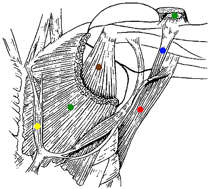
Axillary Arch and Sternalis muscles.
- The axillary arch (blue dot) extends between pectoralis major (green dots) and latissimus dorsi (red dot). Pectoralis minor (brown dot) and a sternalis muscle (yellow dot) are also demonstrated. After Wilder, H.H.: The History of the Human Body, Holt, New York, 1923. From Gehry, K. (1903).
Anatomy Atlases. Immagine ospitata dal sito d’origine. Apri la pagina originale.
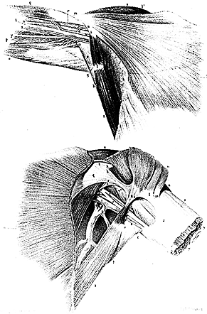
Axillary Arches
from Calori. — Anatomy Atlases. Immagine ospitata dal sito d’origine. Apri la pagina originale.
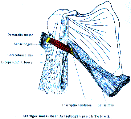
Axillary Arch .
from Tobler. — Anatomy Atlases. Immagine ospitata dal sito d’origine. Apri la pagina originale.
Riferimenti dell’opera originale
Elenco riportato come compare nella fonte, senza normalizzazione dei formati bibliografici.
- Bergman, R.A. (1991) Doubled pectoralis quartus, axillary arch, chondroepitrochlearis, and the twist of the tendon of pectoralis major. Anat. Anz. 173:23-26.
- Birmingham, A. (1889) Homology and innervation of the achselbogen and pectoralis quartus, and the nature of the lateral cutaneous nerve of the thorax. J. Anat. Physiol. 23:206-223.
- Calori, L. (1866) Intorno ad alcune varieta incontrate nella muscolatura degli arti superior. Mem. Accad. Sci. Istituto di Bologna. S.2.,6:157-174.
- Chiaruge, G. (1884) Varietà anatomische. 1.° Varieta muscolare combinata del grande dorsale e del gran pettorale. Bollettino/ Societa tra e Cultoridelle Scienze Mediche in Siena. 2:56-61.
- Dbaly, J. (1975) Ein Fall von beiderseitigem Vorkommen des muskul Achselbogens. Anat Anz. 137:75-78.
- Endres, H. (1893) ein Zwischenmuskelbundel im Gebiete des M. pectoralis major und latissimus dorsi. Anat. Anz. 8:387-397.
- Fritsch, G. (1869) Abnorme Muskelbundel der Achselhöhle. Arch. Anat. Physiol. Wissen. Med. 1869:367-371.
- Gehy, K. (1903) Neue Beiträge zur geschichte des Achselbogens des Menschen, eines Rudimentes des Panniculus der Mammalier. Morphol. Jahrb., Leipzig 31:446-452.
- Huntington, G.S. (1905) The derivation and significance of certain supernumerary muscles of the pectoral region. J. Anat. Physiol. 39:1-54.
- Kaplan, E.B. (1945) Langer's muscle of the axilla. Bull. Hosp. Joint Dis. 6:78-79.
- Kasai, T. and S. Chiba. (1977) True nature of the muscular arch of the axilla and its nerve supply. Kaibogaku Zasshi. 25:657-669.
- Kasai,T and S. Chiba. (1977) True nature of the muscular arch of the axilla and its nerve supply. Kaibogaku Zasshi 52:309-336. In Japanese.
- Kutiyanawala, M.A., Stotter, A., and R. Windle. (1998) Anatomical variants during axillary dissection. Brit. J. Surgery 85:393-394.
- Langer, C. (1846) Zur Anatomie des Musculus latissimus dorsi. Oesterr. Med. Wochenschrift. 1846:454-458, 486-492.
- Lo-Cascio, G. (1915) Contribution a la morphologie de l'arc axillaire de Langer. Arch. Ital. Biol. 62:285.
- Marciniak, T., and K. Serafin. (1929) On the innervation of the axillary arch by the N. intercostobrachialis. Folia Morphologica (Warsaw) 1:23-35.
- Miyauchi, R. (1982) A very rare variation of the latissimus dorsi muscle- a case with accessory insertion of the latissimus dorsi into the first rib and into the pectoralis major muscle. Okajimas Folia Anat. Japan. 58:521-534.
- Monteiro, H. (1926) L'arc axillaire musculaire et ses relations avec les faisceaus pectoreau aberrants. L'Assoc. Anatomistes, Comptes Rendus. 21:262-269.
- Nakajima, K., Chiba, S., Kobayashi, K., Wakatuki, E., Kumaki, K. and T. Hoshino. (1999) A rare muscular anomaly in the upper arm &endash; the chondroepitrochlearis muscle with an aberrant type of the muscular arch of axilla. Kaibogaku Zasshi 74:209-213. In Japanese.
- Pires de Lima, J.A. (1914) Nova série de observacõs Portuguesas de anomalias musculares. Arch. de Anat. e de Anthropol. 1:231-260.
- Phillips, W.F.R. (1913) Innervation of an axillary arch muscle. Anat. Rec. 7:131-132.
- Pichler, K. (1916) Ueber den Langer'schen Achselbogen. Anat. Anz. 49:310-318.
- Pichler, K. (1916) Achelbogen und M. chondro-epitrochlearis bei demselben Trager. Anat. Anz. 49:383-384.
- Pitzorno, M. (1911) Contributo alla morphologia dell'arco ascellare muscolare di Langer. Arch. Ital. Anat. Embriol. 10:129-144.
- Princeteau, -. (1892) Note pour servir a l'historie des anomalies musculaires du creux de l'aisselle. Soc. de Biol., Comptes Rendus Hebdomadaires des Séances et Mémoires 44:202-206.
- Ramsay, A. (1812) An account of unusual conformation of some muscles and vessels. Edinburgh Med. Surg. J. 8:281-283.
- Freiherren von Saar, G. (1903) Zur vergleichenden Anatomie der Brustmuskeln und des Deltamuskels. Arch. f. Anat u. Entwickelungsgeschichte 1903:153-202.
- Sachetello, C.R. (1977) The axillopectoral muscle (Langers axillary arch): a cause of axillary vein obstruction. Surgery 81:610-612.
- Serpell, J.W. and M. Baum. (1991) Signifcance of "Langers axillary arch" in axillary dissection. Australian New Zealand J. Surgery 61:310-312.
- Struthers, J. (1854) On some points in the abnormal anatomy of the arm. British and Foreign Medico-Chirurgical Review, April, 26:523-533, July:224-236.
- Takafugi, T., Igarashi, J., Kanabyashi, T., Yokoyama, T., Moriya, A., Azuma, S. and Y. Sato. (1991) The muscular arch of the axilla and its nerve supply in Japanese adults. Kaibogaku Zasshi 66:511-523. In Japanese.
- Tobler, L. (1902) Der Achselbogen des Menschen, ein Rudiment des Panniculus carnosus der Mammalier. Morph. Jahrbuch 30 (3): 453-507.
- Voto, S.J. and D.S. Weiner. (1987) The chondroepitrochlearis muscle. J. Ped. Orthopedics 7:213-214.
- Weil, -. (1869) Faisceau supplémentaire du grand pectoral s'unissant dans l'aisselle par une expansion aponévrotiqueè un autre faisceau longeant le grand dorsal. Lyon Méd. 1:558-559.
- Weissberg, H. (1932) Über einen Fall von muskulosem Achselbogen. Anat. Anz. 74:105-117.
- Yüksel, M., Yüksel, E. and S. Sürücü. (1996) An axillary arch. Clin. Anatomy 9:252-254.
Classificazione anatomica
- Sistemi:
- Sistema muscolare
Provenienza del contenuto
Adattamento
Adattamento di «Axillary Arch — Illustrated Encyclopedia of Human Anatomic Variation: Opus I: Muscular System» di Ronald A. Bergman, Adel K. Afifi, Ryosuke Miyauchi. Fonte: https://www.anatomyatlases.org/AnatomicVariants/MuscularSystem/Text/A/47Axillary.shtml Opera originale distribuita con licenza CC BY-NC-SA 4.0 (https://creativecommons.org/licenses/by-nc-sa/4.0/deed.it). Adattamento a cura di Redazione AIVA. Modifiche rispetto all’originale: Testo originale inglese riprodotto integralmente e riorganizzato nel formato scheda del portale (sintesi, testo, riferimenti), senza modificarne le affermazioni. La traduzione italiana non è ancora stata eseguita, e la pagina lo dichiara. Le figure non sono state copiate: sono collegate alla pagina d’origine. Questa pubblicazione non è approvata né avallata dagli autori dell’opera originale.
Licenza
Questo contenuto è distribuito con licenza CC BY-NC-SA 4.0. Licenze e attribuzioni