Voce enciclopedica
Axillary Artery
Abstract
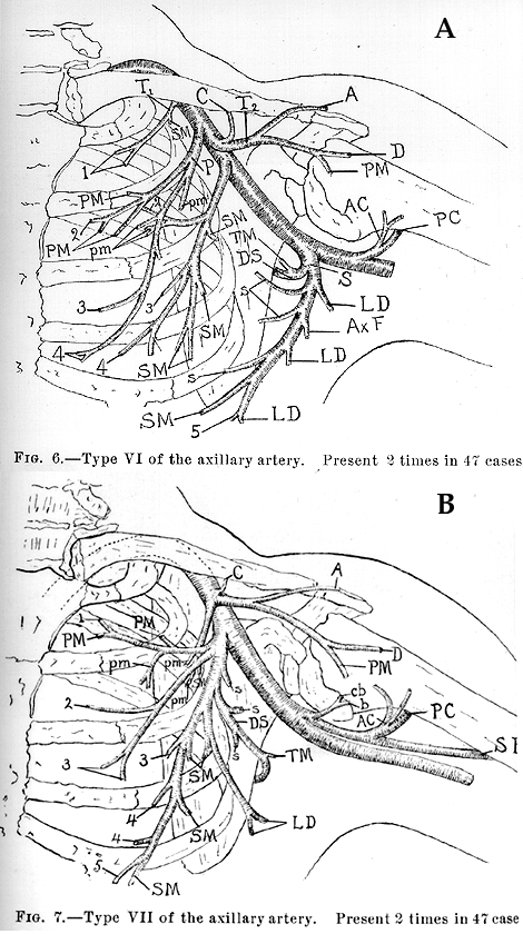
The major variations of the axillary artery are discussed here; there are many! Occasionally, it gives rise to the radial artery or, more rarely, to the ulnar artery. Still more rarely, it gives rise to the interosseous artery or a vas aberrans. It may give rise to a common trunk from which may arise the subscapular, anterior and posterior circumflex humeral, profunda brachii, and ulnar collateral arteries. The branches of the brachial plexus may surround this common trunk, but not the main brachial artery.
The major variations of the axillary artery are discussed here; there are many!
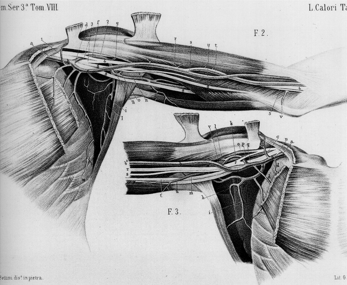
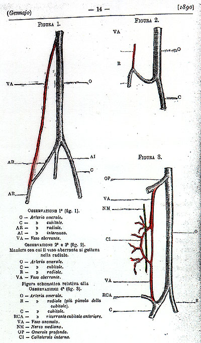
Occasionally, it gives rise to the radial artery or, more rarely, to the ulnar artery. Still more rarely, it gives rise to the interosseous artery or a vas aberrans.
It may give rise to a common trunk from which may arise the subscapular, anterior and posterior circumflex humeral, profunda brachii, and ulnar collateral arteries. The branches of the brachial plexus may surround this common trunk, but not the main brachial artery.
In some cases, the brachial artery appears to be an enlarged "vas aberrans," and the common trunk to the brachial artery, the lower portion of which has been obliterated. A vas aberrans, according to Quain, is a long, unusually slender vessel that originates either from the axillary or the brachial and unites with forearm arteries or their branches. The vas aberrans may extend into the hand to join the superficial palmar arch. In eight of nine cases, the aberrant vessel joined the radial artery; hence it rarely joins the ulnar.
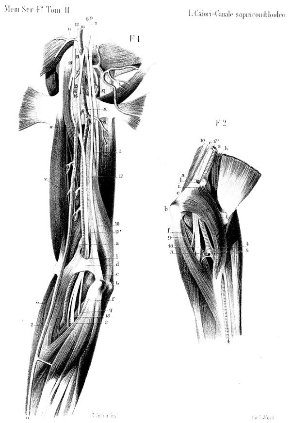
The axillary artery may be represented by two parallel vessels that arise from the first part of the subclavian and continue distally as the ulnar and radial arteries.(Calori)
The first part of the axillary may also provide an accessory thoracoacromial artery. The third part of the axillary artery is occasionally covered by a muscular slip (an axillary arch muscle) derived from the upper part of the tendon of latissimus dorsi; this muscular slip is "always" present in early fetal life, but it usually atrophies later. Axillary arch muscles may persist and may be clinically important. In an article by Alexander Ramsay, Account of unusual conformations of some muscles and vessels, 1813 (see reference list for full citation) the author remarks, " that in 1795, he discovered a transverse muscle of the axilla, which may account for the liability of some subjects to swelling of the axillary glands (nodes) on occasions of violent exertion. This body is composed of an oblong muscle, stretched from the pectoral muscle to the latissimus dorsi and teres major, which in its violent contractions, must prove inconvenient to the axillary arteries, veins, and nerves, which lie beneath this muscle. It was not known in Edinburgh or London, when I demonstrated it, and it is now detected in one of thirty subjects, when dissectors are attentive." The muscle is now known as an axillary arch muscle. It has been well studied by Langer (and others) whose name is often given with it, e.g. Langer's muscle. A more suitable name would be, perhaps, Ramsay's muscle. The Langer citation can be found in Part 1, Muscular System of the present series under Axillary Arch Muscle.
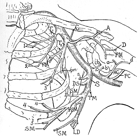
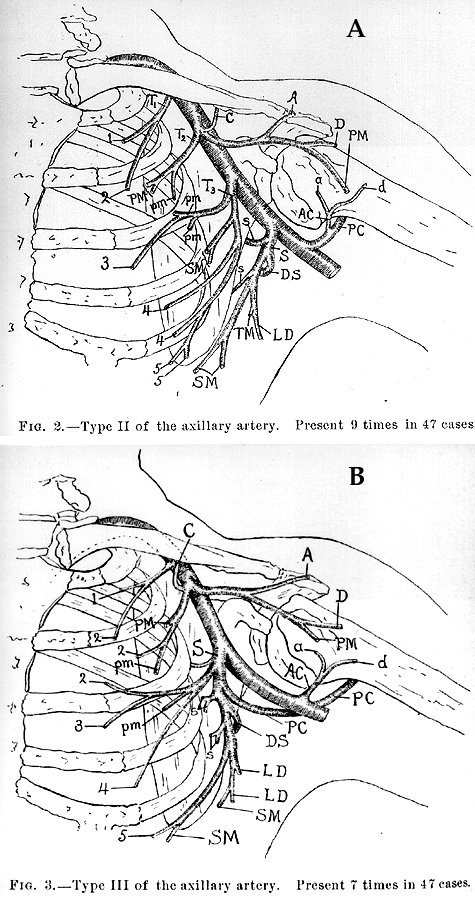
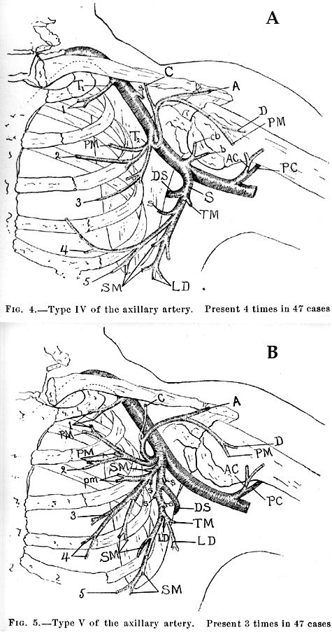
Any of the following branches that normally arise from the subclavian artery may also arise instead, from the axillary artery, either from a common trunk or in groups: the inferior thyroid, ascending cervical, superficial cervical, deep cervical, internal thoracic, transverse cervical, suprascapular or the anterior and posterior circumflex humerale arteries from a common trunk. A trunk from the axillary has been reported giving rise to the anterior and posterior circumflex humeral, the subscapular, the superior profunda, and the inferior profunda arteries (Walsham).
Unusual branches of the axillary include a glandular artery to lymph nodes and skin of the axilla (so called alar thoracic artery) and an accessory lateral thoracic artery.
Heuston reported on three interesting axillary arteries as follows. An aberrant artery, which arose from the axillary, above the origin of the subscapular branch, passed in the arm to the inner (medial) side of the brachial artery, to unite with the radial about two inches below its commencement. In this case the brachial artery bifurcated about one inch (2.54 cm) above its normal position. In a second case, the unusual artery also arose from the axillary, opposite the upper border of teres major muscle, and passing between the heads of the median nerve, proceeded down the outer (lateral) side of the brachial artery, to unite with the radial recurrent, which was large in size in the antecubital fossa. In the third case, the suprascapular artery, arising from the axillary opposite the second rib, passed upwards beneath the clavicle through the cords of the brachial plexus, and having given a branch to the subscapularis muscle, passed beneath the transverse ligament (of the scapula) with the suprascapular nerve, to be distributed as usual.
Konarik, Katchlik, and Baca reported a case with "a coincidental variation of the axillary artery: with a brachioradial artery (radial with a high origin at the end of the axillary artery) and an aberrant posterior humeral circumflex artery passing under the tendon of the latissimus dorsi muscle.
Arteria Aberrans
Anatomy Atlases. Immagine ospitata dal sito d’origine. Apri la pagina originale.
Arteria Aberrans
Anatomy Atlases. Immagine ospitata dal sito d’origine. Apri la pagina originale.
Arteria Aberrans
Anatomy Atlases. Immagine ospitata dal sito d’origine. Apri la pagina originale.
Arteria Aberrans
Anatomy Atlases. Immagine ospitata dal sito d’origine. Apri la pagina originale.
Arteria Aberrans
Anatomy Atlases. Immagine ospitata dal sito d’origine. Apri la pagina originale.
Arteria Aberrans
Anatomy Atlases. Immagine ospitata dal sito d’origine. Apri la pagina originale.
Anatomy Atlases. Immagine ospitata dal sito d’origine. Apri la pagina originale.
Riferimenti dell’opera originale
Elenco riportato come compare nella fonte, senza normalizzazione dei formati bibliografici.
- Anson, B.J., Wright, R.R. and J.A. Wolfer (1939) Blood supply of the mammary gland. Surg., Gynecol. Obstet. 69:468-473.
- Beaumanoir (de Brest), -. (1882) Présentation de pièces anatomiques: axillaire droite et gauche. Bull. de la Soc. Anatomique de Paris 57:318, 320.
- Beaumanoir, (de Brest.), -. (1882) Anomalies artèrielles trouvées a l'autopsie d'un sujet. Bull. de La Soc. Anatomique de Paris 57:316-321.
- Bergman, R.A., Thompson, S.A., Afifi, A.K. and F.A. Saadeh. (1988) Compendium of Human Anatomic Variation: Catalog, Atlas and World Literature. Urban & Schwarzenberg. Baltimore and Munich.
- Bhargava, I. (1956) Anomalous branching of axillary artery. J. Anat. Soc. India. 5:78-80.
- Bovero, A. (1907) Morphologie des artères de la glande mammaire. Arch. Ital. Biol. 48:486-487.
- Calori, L. (1877) Sul variato rapporto vascolare delle due radici del nervo mediano e sulle condizioni atte a produrlo. Mem. Accad. Sci. Instituto di Bologna S. 3. 8:443-456.
- Cayotte, J. and P. Quereux (1953) Ramescence anormale des artères axillaire et fémorale. Assoc. Anatomistes Comptes Rendus. 40:968-972.
- Coulouma, B. (1934) Resultat de cent quatre observations su la disposition des branches de l'axillaire. Bull. Assoc. Anat. (Nancy). 29:193-199.
- Ferrari, G. (1902-03) Anomalie di rapporti e distribuzione dell' arteria ascellare. Atti della Accademia delle scienze mediche e naturali in Ferrara 77:259-262.
- Galinos, -. et P. Farabeuf. (1907) Anomalie des artères du membre supérieur. Bull. et Mém. de la Soc. Anatomique de Paris 9:153-155.
- DeGaris, C.F. and W.B. Swartley. (1928) The axillary artery in white and negro stocks. Am. J. Anat. 41:353-397.
- Guerra, P. (1887) Anomalie rinvenute in cadaveri di delinquenti e di normale. Ateneo Medico Parmense 1:278-292.
- Heuston, F.T. (1885) Notes on some anatomical anomalies. Transactions - Royal Academy of Medicine in Ireland 3:453-456.
- Hitzrot, J.M. (1901) A composite study of the axillary artery in man. Johns Hopkins Hosp. Bull. 12:136-145.
- Huelke, D.F. (1959) Variation in the origins of the branches of the axillary artery. Anat. Rec. 135:33-41.
- Jakubowicz, M. and M. Bruska. (1980) A case of variations in arterial vascularization of the upper limb. Folia Morphologica, Warsaw 39:55-59.
- Josso, -. (1879) Anomalie des artères du bras. Bull. de la Société Anatomique de Nantes 1879:10-11.
- Konarik, M., Katchlik, D., and V. Baca. (2014) A coincidental variation of the axillary artery: the brachioradial artery and the aberrant posterior humeral circumflex artery passing under the tendon of the latissimus dorsi muscle. Bos J Basic Med Sci 2014:1995-2014.
- Kubo, -. and -. Shirota (1906) Varietäten der Aeste der A. axillaris. In Japanese. Zasshi/ Tokyo Igakkai 20:467-481.
- Mauclaire, -. (1895) Dedoublement de l'artere axillaire avec plusieurs rameaux nerveux au niveau de l'angle de bifurcation. Bull. et Mém. de la Soc. Anatomique de Paris. 9:135-136.
- Miller, R.A. (1939) Observations upon arrangement of the axillary artery and brachial plexus. Am. J. Anat. 64:143-163.
- P'an, Ming-Tzu. (1940) The origin of the branches of the axillary artery in the Chinese. Am. J. Physical Anthropol. 27:269-279.
- Parmentier, -. (1850) Anomalies artérielles; l'artère axillaire. Bull. et Mém. de la Soc. Anatomique de Paris 1850:74-75.
- Pellegrini, A. (1904) Il tipo normale e le variazioni delle Arteriae subclavia e axillaris. Monitore Zoologico Italiano 15:232-244.
- Pellegrini, A. (1906) Le arteriae subclavia e axillaris nell'uomo studiate col metodo statistico. Arch. Ital. Anat. Embriol. 5:205-255, 466-505.
- Petrequin, -. (1837) Artère humérale double par suite de la bifurcation de l'artère axillaire.Gazette Médicale de Paris 5:199.
- Potherat, E. (1888) Anomalie de l'axillaire. Bull. et Mém. de la Soc. Anatomique de Paris 1888:296-298.
- Poynter, C.W.M. (1922) Congenital anomalies of the arteries and veins of the human body with bibliography. The University Studies of the University of Nebraska. 22:1-106.
- Ramsay, A. (1813) Account of unusual conformations of some muscles and vessels. Edinburgh Medical and Surgical Journal 8:281-283.
- Schwalbe, E. (1898) Beitrag zur Kenntniss der Arterienvarietäten des menschlichen Arms. Morphologische Arbeiten 8:1-47.
- Skopakoff, C. (1959) Uber die Variabilität der Abzweigung der Hauptäste der Aa. axillaris et brachialis. Anat. Anz. 107:294-304.
- Swensson, G. och P. Wetterdal. (1916) Armartärena hos människan. Svenska Lakaresallskapets Handlingar. Acta Societatis Medicorum Suecanae 1916:1319.
- Trotter, M., Henderson, J.L., Gass, H., Brua, R.S., Weisman, S., Agress, H., Curtis, G.H. and E.R. Westbrook. (1930) The origins of the branches of the axillary artery in Whites and American Negroes. Anat. Rec. 46:133-137.
- Walsham, W.J. (1880) Anatomical variations: An account of a few of the more interesting abnormalities that have occurred in the dissecting-rooms during the past seven years; with remarks on their morphological significance, and their bearing on the practice of surgery. St. Bartholomews Hospital Reports 16:91.
- Winslow, R. (1883) A study of the malformations, variations, and anomalies of the circulatory apparatus in man. Annals of Anatomy and Surgery 7:18-19.
- Zagorsky, P. (1809) Observationum anatomicarum quadrigae de singulari arterium aberratione. Mem. de l'Acad. Imperiale des Sciences de St. Petersbourg 1:386.
- Variations in Branches of Axillary Artery The following variations have been recorded in the branches of the axillary artery.
- The first part may give rise to the dorsal (posterior) scapular artery (0.5% of individuals, Poynter). Pellegrini found the first branch to be one to serratus anterior (20% of individuals) and Poynter found this same vessel in 42% of 200 cases.
Classificazione anatomica
- Sistemi:
- Sistema cardiovascolare
Provenienza del contenuto
Adattamento
Adattamento di «Axillary Artery — Illustrated Encyclopedia of Human Anatomic Variation: Opus I: Muscular System» di Ronald A. Bergman, Adel K. Afifi, Ryosuke Miyauchi. Fonte: https://www.anatomyatlases.org/AnatomicVariants/Cardiovascular/Text/Arteries/Axillary.shtml Opera originale distribuita con licenza CC BY-NC-SA 4.0 (https://creativecommons.org/licenses/by-nc-sa/4.0/deed.it). Adattamento a cura di Redazione AIVA. Modifiche rispetto all’originale: Testo originale inglese riprodotto integralmente e riorganizzato nel formato scheda del portale (sintesi, testo, riferimenti), senza modificarne le affermazioni. La traduzione italiana non è ancora stata eseguita, e la pagina lo dichiara. Le figure non sono state copiate: sono collegate alla pagina d’origine. Questa pubblicazione non è approvata né avallata dagli autori dell’opera originale.
Licenza
Questo contenuto è distribuito con licenza CC BY-NC-SA 4.0. Licenze e attribuzioni