Voce enciclopedica
Brachial Artery
Abstract
Major variations are present in about 25% of subjects studied and Poynter states that the variations are all explainable on an embryological basis. Classification of Arm Arterial Variations (Müller, 1903, from Poynter). Double brachial, i.e., superficial and deep: Superficial brachial continues as the: Superficial brachial as the radial and superficial antibrachial. In Group I.A. , the brachial is doubled and consists of deep and superficial branches. It is the homologue of the superficial brachial described by Bayer (1893) for monkeys. Haller called it vasa aberrentia.
Major variations are present in about 25% of subjects studied and Poynter states that the variations are all explainable on an embryological basis.
Classification of Arm Arterial Variations (Müller, 1903, from Poynter).
- Brachial artery passes in front of the median nerve and continues as:
- Arteria brachialis superficialis superior.
- Arteria brachialis inferior.
Double brachial, i.e., superficial and deep:
- Brachialis superficialis superior.
- Brachialis inferior.
- Superficial and deep reunite.
- Superficial continues as the radial,
- without anastomosis.
- with anastomosis.
Superficial brachial continues as the:
- Ulnar,
- Median,
- Ulnar and Median.
Superficial brachial as the radial and superficial antibrachial.
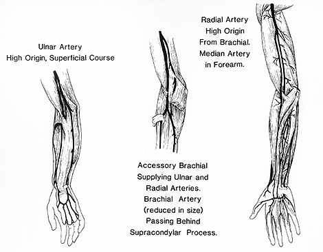
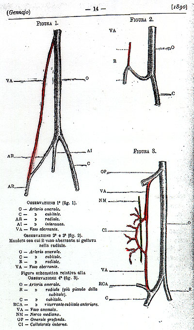
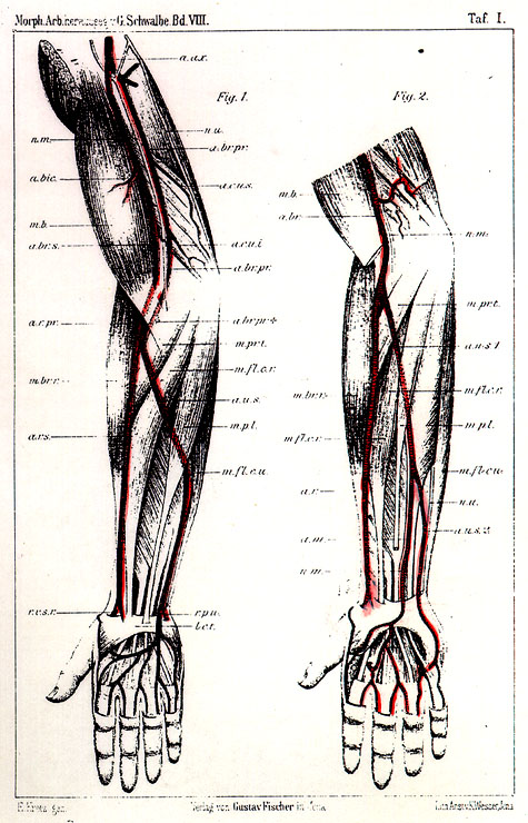
In Group I.A. , the brachial is doubled and consists of deep and superficial branches. It is the homologue of the superficial brachial described by Bayer (1893) for monkeys. Haller called it vasa aberrentia. In these cases, when the superficial branch persists it terminates in the proximal part of the radial, the ulnar, or the distal part of the brachial trunk. This variation was first reported by Ludwig in 1767.
Group I.B. , The profunda drops out and the superficial brachial persists as the single trunk in the arm. This variation was reported by Chassaignac in 1834.
Group II.A. , The brachial is doubled and the superficial brachial continues as the radial. This is known as high origin of the radial and is the most common of all arm variations. Müller has shown that developmentally the superficial brachial arises in three parts and therefore can account for the variation in the origin extending from the axillary to the lower third of the arm. Variations of this type were seen and reported by Fabricius (1754), Eschenbach (1755) and by Morenheim (1783).
Group II.B. , The brachial is doubled and the superficial brachial continues as the ulnar.
Group II.1. , The brachial is doubled and the superficial brachial continues as the median or the interosseus. In these cases the usual brachial continues as the radial and ulnar in the usual way. Less common than the preceeding two groups the variation represents the dropping out of the connection between the ulnar and the median. First noted by Barclay in 1812.
Group II.2. , In this group the brachial is doubled and the superficial brachial continues as the radial and ulnar, the profunda as the interosseus. Probably first reported by Meckel in 1816.
Group II.3. , The superficial antibrachial radial may be present either as a continuation of the superficial brachial or from profunda brachii. Found by Dubrueil in 1847.
Group II.4. , Superficial antibrachial persists, ulnar may be doubled. This condition was described by Lucas in 1838.
In one study of 610 limbs, the usual textbook description of the axillary continuing as the brachial was observed in 80% of specimens.
A superficial brachial, a vessel that has a course anterior to that of the median nerve, was found in about 13%. The superficial branch continued as the radial twice as frequently as it continued as the ulnar artery. Less frequently it continued as both arteries.
A communication at the elbow between the superficial brachial and the usual brachial was seen occasionally.
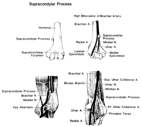
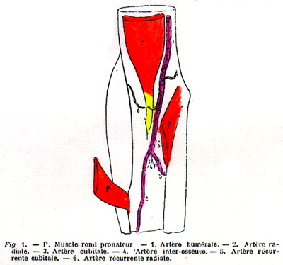
One of the major variations is a high proximal division into terminal branches (radial, 15%), ulnar (2%), common interosseous, vas aberrans, and superficial median antebrachial artery). This may occur at any point in the normal course of the vessel, but is most common in the upper third of the arm and least common in the middle third. The two vessels into which the brachial divides run parallel to each other to the bend of the elbow, in the usual position of the brachial. From this point, one branch follows the normal course of the radial artery through the forearm and the other takes the normal course of the ulnar artery, supplying as usual the common interosseous artery. This arrangement is considered a simple high division of the brachial artery. In the following cases, the disposition of the two vessels is different: 1) the two arteries communicate at the elbow by a cross branch, or reunite and then divide again in the usual manner. 2) one vessel follows the course of the ulnar artery in the forearm, and the other divides into the radial and common interosseous. 3) one artery divides into the radial and ulnar as usual, and the other takes the course of the common interosseous and divides into the anterior and posterior interosseous arteries or, much more rarely, takes the course of the posterior interosseous artery. The anterior interosseous comes from the ulnar and finally, 4) the vessels follow a different course in the upper arm different from that of the usual brachial.
The branch representing the radial may: a) cross over or under the other branch, b) perforate the deep fascia above the elbow and run beneath the skin to its place in the forearm, or c) pass behind the tendon of the biceps.
The branch representing the ulnar may: 1) run to the front of the medial epicondyle with the median nerve, reaching its usual position by descending beneath the fascia and the pronator teres muscle or, more rarely, beneath some of the flexor muscles or merely beneath the skin, or 2) pass with the ulnar nerve behind the medial epicondyle and beneath the muscles to its usual place in the forearm.
An enlarged vas aberrans (Haller) may be present. This long vessel arises from the brachial, usually near the origin of the profunda, and joins, most commonly, the radial artery or, more rarely, one of its branches or the ulnar. Normally it is quite small; it descends over the median nerve to the biceps muscle. At times this vessel takes the place of the brachial; the median nerve will then be found behind the artery.
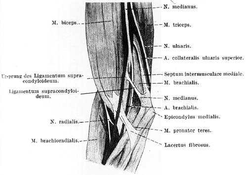
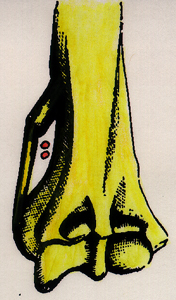
The brachial may run with the median toward the medial epicondyle, where it may turn around, or beneath, a suprcondylar process if present (2.7% of individuals, Gruber) and then descend to its normal position beneath the pronator teres. It may also pass through the pronator teres muscle where it may be entrapped and compromised.
Latarjet suggests five possibilities for branching from a premature division of the brachial artery: 1) Radial and ulnar-common interosseous trunk; 2) Ulnar and a radial-common interosseous trunk; 3) Common interosseous or persistent median artery and a radioulnar trunk; 4) Radial, ulnar, and a common interosseous artery and 5) A vas aberrans and normal brachial artery.
The variations of this vessel is usually unilateral. Quain reported 61 cases, with 43 involving one side only and 18 involving both sides. In five of the 18 cases, the same variation was bilateral, and in 13 cases the variation differed on the two sides.
The brachial artery may give rise to a median cubital, a median articular cubital artery, or a branch to the mammary gland. It may be covered by various muscular slips derived from the adjacent muscles or those in its course.
The brachial artery may be compressed by the lacertus fibrosus of the biceps brachii muscle.
The frequency of brachial variations based on studies by various authors (3337 upper limbs) is about 20%.
Deep (Profunda)
Anatomy Atlases. Immagine ospitata dal sito d’origine. Apri la pagina originale.
Deep (Profunda)
Anatomy Atlases. Immagine ospitata dal sito d’origine. Apri la pagina originale.
Deep (Profunda)
Anatomy Atlases. Immagine ospitata dal sito d’origine. Apri la pagina originale.
Deep (Profunda)
Anatomy Atlases. Immagine ospitata dal sito d’origine. Apri la pagina originale.
Deep (Profunda)
Anatomy Atlases. Immagine ospitata dal sito d’origine. Apri la pagina originale.
Deep (Profunda)
Anatomy Atlases. Immagine ospitata dal sito d’origine. Apri la pagina originale.
Deep (Profunda)
Anatomy Atlases. Immagine ospitata dal sito d’origine. Apri la pagina originale.
Deep (Profunda)
Anatomy Atlases. Immagine ospitata dal sito d’origine. Apri la pagina originale.
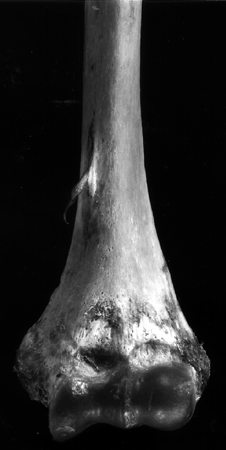
Deep (Profunda)
Anatomy Atlases. Immagine ospitata dal sito d’origine. Apri la pagina originale.
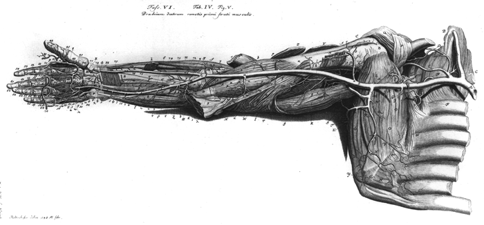
Deep (Profunda)
Anatomy Atlases. Immagine ospitata dal sito d’origine. Apri la pagina originale.
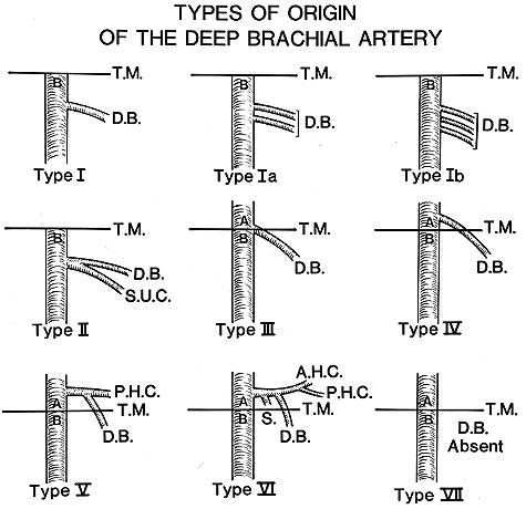
Superficial inferior
Anatomy Atlases. Immagine ospitata dal sito d’origine. Apri la pagina originale.
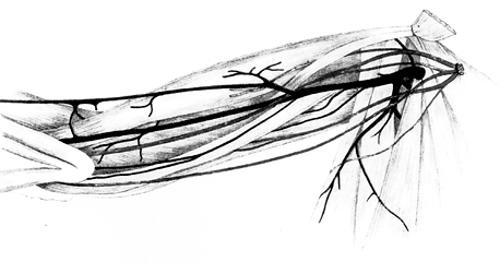
Superficial superior
Anatomy Atlases. Immagine ospitata dal sito d’origine. Apri la pagina originale.
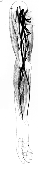
Anatomy Atlases. Immagine ospitata dal sito d’origine. Apri la pagina originale.
Riferimenti dell’opera originale
Elenco riportato come compare nella fonte, senza normalizzazione dei formati bibliografici.
- Adachi, B. (1928) Das Arteriensystem der Japaner. Bd. 1 Verl. d. Kaiserl. - Jap. Univ., Kyoto.
- Anson, B.J., Ed. (1966) Morris' Human Anatomy, 12th ed., The Blakiston Division, McGraw-Hill Book Company, New York.
- Barta, E. and L. Petrovits. (1927) Das Ligamentum supracondyloideum. Anat. Anz. 63:177-184.
- Bassett, III., F.H., Spinner, R.J. and T.A. Schroeter. (1994) Brachial artery compression by the lacertus fibrosus. Clinical Orthopaedics 307:110-116.
- Bayer, L. (1893) Beitrag zur vergleichenden Anatomie der Oberarmarterien. Morph. Jahrbuch 19:1-41.
- Beaumanoir (de Brest), -. (1882) Présentation de pièces anatomiques:humérale droite. Bull. de la Soc. Anatomique de Paris 57:318-319.
- Beaumanoir, (de Brest.), -. (1882) Anomalies artèrielles trouvées a l'autopsie d'un sujet. Bull. de La Soc. Anatomique de Paris 57:316-321.
- Bergman, R.A., Thompson, S.A., Afifi, A.K. and F.A. Saadeh. (1988) Compendium of Human Anatomic Variation: Catalog, Atlas and World Literature. Urban & Schwarzenberg., Baltimore and Munich.
- Bertacchini, P. (1896) Un cas d'anomalie artérelle du bras et d'apophyse susépitrochléenne de l'humérus associés. Arch. Ital. Biol. 26:159.
- Bianchi, L. (1943) Considerazioni sopra un caso di arteria brachio-ulnare superficiale. Anat. Anz. 94:73-88.
- Bonora, C. e L. Casati. (1880) Anomalia dell'Arteria omerale ed aneurisma della piegatura del braccio. Raccoglitore Medico 13:175-188.
- Bourguet, -. (1905) Autra cas d'anomalies artérielles du membre supérieur. Tolouse Mddical Juin 15,7(12):133-136.
- Brême, G. (1899) Casuistischer Beitrag zur Kenntniss der Anomalien der Armarterien. Zeitschr. für Morphologie und Anthropologie 1:483-494.
- Broca, -. (1850) L'artérielle humérale. Bull. et Mém. de la Soc. Anatomique de Paris 1850:75-76.
- Broca, -. (1849) Cinq anomalies artérielles sur le même subject. Anomalies rare des artères de l'avant--bras. Reflexions sur les anomalies artérielles du member thoracique (popliteal, renales, deux arteres humérals, femoral, tibio-peronier tronc). Bulletins et Mem. de la Société Anatomique de Paris XXIV(2):49-59.
- Brunati, J. (1937) Sur les bifurcations hautes de l'artère humerale. Ann. Anat. Path., Paris 14:873-882.
- Bussière, -. (1904) Sur quelques anomalies artérielles. Journal de Medecine de Bourdeaux Février, 1904) 6:101-102.
- Cappi, E. (1879) Di un'anomalia della arterie omerali. Annali Universali di Medicina e Chirurgia 47:267-268.
- Carson, E.R. (1895) Rare anomaly of the brachial artery. Medical Record 48:911-
- Charles, C.M., Penn, L., Holden, H.F., Miller, R.A. and E.B. Alvis. (1931) The origin of the deep brachial artery in American white and American negro males. Anat. Rec. 50:299-302.
- Charles, J.J. (1873) Notes on some cases of abnormal arrangement of arteries of the upper extremity. J. Anat. Physiol. 7:300-301.
- Charles, J.J. (1875) Abnormalities in the arteries of the upper extremity. J. Anat. Physiol. 9:180-181.
- Clavier, -. (1873) Bifurcation prématurée de l'artère humerale et double anastomose au pli du coude. Lyon Méd. 12:457.
- Clavier, -. et -. Boliet. (1873) Dualitè de l'artère humérale, variété huméro-cubitale; noyaux osseux à l'articulation du coude. Lyon Méd. 12:387.
- Cocchi, A. (1891) Contribuzione allo studio dell'anastomosi tra radiale e cubitale alla piegatura dell cubito nella divisione prematura dell'arteria brachiale. Atti dell Accademia dei Fisiocritici in Siena. Ser.4, 3:247-261.
- Corson, E.R. (1895) A rare anomalie of the brachial artery. The Medical Record, N.Y. 48:911.
- Cruvielhier, -. (1834) Anomalie de l'artère brachiale. Bull. et Mém. de la Soc. Anatomique de Paris 1834:40-41.
- Cruveilhier, -. (1835) Deux cas d'anomalie dans la distribution de l'artère brachiale. Bull. et Mém. de la Soc. Anatomique de Paris 1835:2.
- Curtet, -. (1874) Artère huméro-radiale aberrante et anomalie du biceps. Lyon Méd. 16:162.
- Dean, M.H. (1880) Arterial Anomalies. The Medical Record, N.Y. 17:384.
- Dieulaufe, R. et M. Cahuzac. (1931) Un cas d'anomalies artèrielles due membre superieur. Ann. Anat. Path., Paris 8:661-662.
- Dubrevil-Chambardel, L. (1926) Variations des Artéres du Membre Supérieur., Masson Cie., Paris.
- Duchamp, -. (1874) Anomalie de l'artère humérale et du nerf médian. Lyon Méd. 16:162.
- Deville, -. (1849) Canal ou conduit sus-épitrochléen (sus-épicondylien des animaux) donnant anormalement passage à l'artère humeral et au nerf median. Bulletins et Mem. de la Société Anatomique de Paris XXIV(7):213-214.
- Esquerdo, A. (1887) De algunas anomalías arteriales. Archivo de La Cirugía 1:86-89.
- Foltz, -. (1871) Dualité de l'artère humérale. Ann. de la Société de Médecine de Lyon Ser.2, 19:145-156.
- Foltz, -. (1872) Anomalie par dualité de l'artère humérale. Lyon Méd. 9:150-160.
- Forster, A. (1917-18) über zwei Fälle einer seltenen Arterienvarietät an der Ansatzsehne des Biceps brachii. Anat. Anz. 50:193-194.
- Georgievski, I.V. (1905) K kazuistik vysokogo proischozdenija i podkoznago polozenija tyl'noj v-tvi lucevoj arteri. Chirurgija 18:319-317.
- Gérard, G. et E. Breucq. (1905) Sur un cas de bifurcation précoce de l'artère humérale. Bibliographie Anatomique 14:164-170.
- Göppert, E. (1904) Die Beurteilung der Arterienvarietaten der oberen Gliedmasse bei den Säugetieren und beim Menschen auf entwicklungsgeschichtlicher und vergleichend-anatomischer Grundlage. Erg. Anat. Entw'gesch. 14:170-233.
- Göppert, E. (1913) Blutgefässsystem. In Gegenbaur, Lehrbuch der Anatomie des Menschen.
- Henle, J. (1868) Handbuch der systematischen Anatomie des Menschen. Von Friedrich Vieweg und Sohn., Braunschweig.
- Herrington, M. (1905) High bifurcation of the brachial artery with reunion at the elbows. Johns Hopkins Hosp. Bull. 16:65-66.
- Hofer, K. and G. Hofer. (1910) über den Verlauf der Arteria brachialis mit dem Nervus medianus zwischen den beiden Köpfen des Musculus pronator teres. Anat. Anz. 36:510-514.
- Humbert, -. (1873) Anomalie de l'artère humérale. Bull. et Mém. de la Soc. Anatomique de Paris 1873:261.
- Jakubowicz, M. and M. Bruska. (1980) A case of variations in arterial vascularization of the upper limb. Folia Morphologica, Warsaw. 39:55-59.
- Janosík, J. (1891) Cérvy krevni a nervy horni koncetiny u cloveka a nekterych zvirat. (Sur les vaisseaux sanguins et les nerfs des membres supérieurs l'homme et chez quelques autres animaux). Sbornik Lekarsky 4:244-255.
- Jaschtschinski, S.N. (1897) Morphologie und topographie des Arcus volaris sublimis und profundus des Menschen. Anat. Hefte (Abt. 1) 7:161-188.
- Jurjus, A., Sfeir, R. and R. Bezirdjian. (1986) Unusual variation of the arterial pattern of the human upper limb. Anat. Rec. 215(1):82-83.
- Kadanoff, D. und G. Balkansky. (1966) Zwei Fälle mit seltenen Varietäten der Arterien der oberen Extremität. Anat. Anz. 118:289-296.
- Krause, E. (1876) Varietäten des Aortensystems. In J. Henle, Handbuch der systematischen Anatomie des Menschen. Bd. 3. Abt. 1.2. Von Friedrich Vieweg und Sohn, Braunschweig.
- Lafaye, (1873) Dualitè de l'artère humérale, variété huméro-radiale, origin d'une radio-palmaire au pli du coude. Lyon Méd. 12:387.
- Lafite-Dupont, -. (1898) D'anomalies diverses de l'artère humérale. Gazette Hebdomadaire des Sciences Médicales de Bordeaux 19:116-117.
- LaGrange, -. (1882) Anomalie de l'artère humérale. Bull. et Mém. de la Soc. Anatomique de Paris 1882:340-341.
- Langer, -. (1851) Varietät der Arter. brachialis. Zeitschr. der Gesellschaft der Aerzte in Wien. 74:185.
- Langer, C. von. (1911) Lehrbuch der systematischen und topographischen. Anatomie. Bd. 1, T.3, S.520 von C. Toldt, Braumüller, Wien - Leipzig
- Lanz,T. von und W. Wachmuth. (1959) Praktische Anatomie. 2. Auf. Bd. 1, T. 3, S.200. Springer, Berlin- Göttingen-Heidelberg.
- Latarjet, A. (1948) Testut's Traite d'Anatomie Humaine, 9th ed., G. Doin & Cie., Paris.
- Loetzke, H.H. and W. Kleinau (1968) Gleichzeitiges Vorkommen der Aa. brachialis superficialis, radialis und antebrachialis dors. superfic. sowie deren Aufzweigungen. Anat. Anz. 122:137-141.
- Maestre, A. (1864) Notable anomalía de la arteria humeral. La España Médica 9:330-331.
- Maruyama, K. (1944) Seltene Varietät der Arterien der oberen Extremität bei einem Japaner (A. brachialis superficialis lateralis inferior, A. antebrachialis superficialis mediano-ulnaris, Arcus volaris superficialis vom Typus mediano-ulnaris). Okajima's Folia Anat. Jpn. 22:551-567.
- McCormack, L.J., Cauldwell, E.W. and B.J. Anson. (1953) Brachial and antebrachial arterial patterns. Surg., Gynecol. Obstet. 96:43-54.
- Meckel, J.F. (1816) Ueber den regelwidrigen Verlauf der Armpulsadern. Dtsch. Arch. Physiol. 2:117-131.
- Müller, E. (1903) Beiträge zur Morphologie des Gefässsystems. 1. Die Armarterien des Menschen. Anat. Hefte Abt.1,22:379-574.
- Pabst, R. and H. Lippert. (1968) Beiderseitiges Vorkommen von A. brachialis superficialis, A. ulnaris superficialis und A. mediana. Anat. Anz. 123:223-226.
- Parmentier, -. (1850) Anomalie de la carotid et de l'artère humérale. Bulletins et Mem. de la Société Anatomique de Paris XXV(3):74-75.
- Petit, -. (1867) Anomalies des artères et des veines du bras. Bulletin Médical du Nord de la France Publié par la Société Centrale de Médecine du Dèpartement du Nord 8:237-242.
- Peterquin, -. (1837) Artère humérale double par suite de la bifurcation de l'artère axillaire. Gazette Médicale de Paris 5:199.
- Peyronny, -. (1902) Anomalies arterielles multiples des arteres du bras. J. de Medecine de Bordeaux et du Sud-Ouest. 32:411-412.
- Polonskaja, R. (1932) Zur Frage der Arterienanastomosen im Gebiete der Ellenbogenbeuge des Menschen. Anat. Anz. 74:303-317.
- Poynter, C.W.M. (1922) Congenital anomalies of the arteries and veins of the human body with biblography. The University Studies of the University of Nebraska. 22:1-106.
- Rauber, -. und -. Kopsch. (1948) Rauber-Kopsch Lehrbuch und Atlas der Anatomie des Menschen. Bd 2., S.444. Theime, Leipzig.
- v. Reinhardt, -. (1856) Beobachtung eines interessanten Falles von Abweichung der Art. brachialis vom normalen Verlauf. Zeitschrift für Wundärzte und Geburtshelfer 9:275-278.
- Robin, -. (1874) Bifurcation de l'artère humérale, donnant une humero-radiale. Lyon Méd. 16:162.
- Rossi, U. (1890) Anomalie Arteriose. Lo Sperimentale, Giornale Italiano di Scienze Mediche 65:3-14.
- Ruge, G. (1884) Beiträge zur Gefässlehre des Menschen. Gegenbaurs Morphologisches Jahrbuch 9:329-388.
- Sandifort, Ed. (1777) Brachiales arteriae binae ex subclavia enatae p. 127. Observationes Anatomico-Pathologicae, Apud P.v.d. Eykk et D. Vygh, Leiden.
- Schwalbe, E. (1889) Beitrag zur Kenntniss der Arterienvarietäten des menschlichen Arms. Morphologichen Arbeiten 8:1-47.
- Schwalbe, E. (1898) Beitrag zur Kenntniss der Arterienvarietäten des menschlichen arms. Morphologische Arbeiten 8:1-48.
- Schaefer, E.A., Symington, J. and T.H. Bryce. (1915) Quain's Anatomy, 12 th ed. Longmans, Green and Company, London.
- Shepherd, F. J. (1883) On some anatomical variations with remarks on their morphology. Annals of Anatomy and Surgery 8:175.
- Siegbauer, F. (1940) Lehrbuch der normalen Anatomie des Menschen. Urban & Schwarzenberg, Wien-Berlin.
- Skopakoff, C. (1959) über die Variabilität der Ab- und Verzweigung der A. brachialis superficialis. Anat. Anz. 106:356-368.
- Skopakoff, C. (1959) über die Variabilität der Abzweigung der Hauptäste der Aa. axillaris et brachialis. Anat Anz. 107:294-304.
- Souligoux, -. (1893) Anomalie de l'artère humérale. Bull. et Mém. de la Soc. Anatomique de Paris 7:278.
- Struthers, J. (1848) On a peculiarty of the humerus and humeral artery. Month. J. Med. Sci. (new series 28) 8:264-267.
- Tédénat, -. (1873) Humerale double, l'une donnant la radiale, l'autre e passant sous une arcade osteo-fibreuse. Lyon Méd. 12:386.
- Tedeschi, -., Torraca, -. and -. Pavone. (1907) Anomalia della arterie dell'antibraccio e del nervo mediano. Giornale Internazionale delle Scienze Mediche 29:315-317
- Vinon, -. et -. Ducos. (1912) Anomalie de l'artère humérale avec apophyse sus-épitrochléenne. Montpellier Médical 30:132.
- Walsham, W.J. (1880) A large branch from the brachial artery distributed as the ulnar in the palm; the ulnar in the forearm derived from the brachial as usual. St Bartholomews Hospital reports 16:91-92.
- Walther, -. (1886) Division tardive de l'artère humérale. - Artère radiale passant au dessus du rond pronateur. Bull. et Mém. de la Soc. Anatomique de Paris 1886:204-205.
- Weathersby, H.T. (1956) Anomalies of brachial and antebrachial arteries of surgical significance. South. Med. J. 49:46-49.
- Winslow, R. (1883) A study of the malformations, variations, and anomalies of the circulatory apparatus in man. Annals of Anatomy and Surgery 7:19-21.
- Yucel, H.H. (1999) Unilateral variation of the arterial pattern of the upper extremity with a muscle variation of the hand. Acta Med. Okayama. 53(2): 61-65.
- Zuckerkandl, E. (1895) Zur Anatomie und Entwicklungsgeschicte der Arterien des Vorderarmes. Anat. Hefte 4:1-98, 5:157-205.
- Variations in Branches of Brachial Artery The following variations have been recorded in the branches of the brachial artery. Profunda Brachii Artery Superior Ulnar Collateral Artery Inferior Ulnar Collateral Artery Ulnar Artery
Classificazione anatomica
- Sistemi:
- Sistema cardiovascolare
Provenienza del contenuto
Adattamento
Adattamento di «Brachial Artery — Illustrated Encyclopedia of Human Anatomic Variation: Opus I: Muscular System» di Ronald A. Bergman, Adel K. Afifi, Ryosuke Miyauchi. Fonte: https://www.anatomyatlases.org/AnatomicVariants/Cardiovascular/Text/Arteries/Brachial.shtml Opera originale distribuita con licenza CC BY-NC-SA 4.0 (https://creativecommons.org/licenses/by-nc-sa/4.0/deed.it). Adattamento a cura di Redazione AIVA. Modifiche rispetto all’originale: Testo originale inglese riprodotto integralmente e riorganizzato nel formato scheda del portale (sintesi, testo, riferimenti), senza modificarne le affermazioni. La traduzione italiana non è ancora stata eseguita, e la pagina lo dichiara. Le figure non sono state copiate: sono collegate alla pagina d’origine. Questa pubblicazione non è approvata né avallata dagli autori dell’opera originale.
Licenza
Questo contenuto è distribuito con licenza CC BY-NC-SA 4.0. Licenze e attribuzioni