Voce enciclopedica
Circle of Willis
Abstract
This circle of anastomosing arterial vessels is extremely variable. Stehbens regards a normal circle of Willis to be one in which there is a complete anastomotic polygon, which may show considerable variability not only of its components but also its branches. The so-called normal or textbook description holds true in about 34.5% of cases based on a composit review of 1413 brains. Not even Willis's original illustration conforms to the usual textbook description. See illustrations for Circle variations.
This circle of anastomosing arterial vessels is extremely variable.
Stehbens regards a normal circle of Willis to be one in which there is a complete anastomotic polygon, which may show considerable variability not only of its components but also its branches. The so-called normal or textbook description holds true in about 34.5% of cases based on a composit review of 1413 brains. Not even Willis's original illustration conforms to the usual textbook description.
See illustrations for Circle variations.
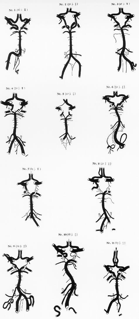
Anatomy Atlases. Immagine ospitata dal sito d’origine. Apri la pagina originale.
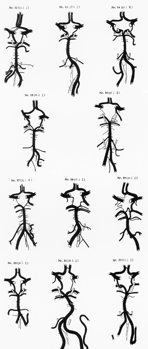
Anatomy Atlases. Immagine ospitata dal sito d’origine. Apri la pagina originale.
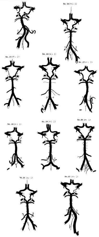
Anatomy Atlases. Immagine ospitata dal sito d’origine. Apri la pagina originale.
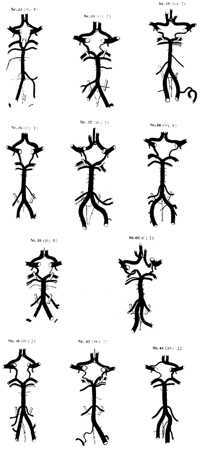
Anatomy Atlases. Immagine ospitata dal sito d’origine. Apri la pagina originale.
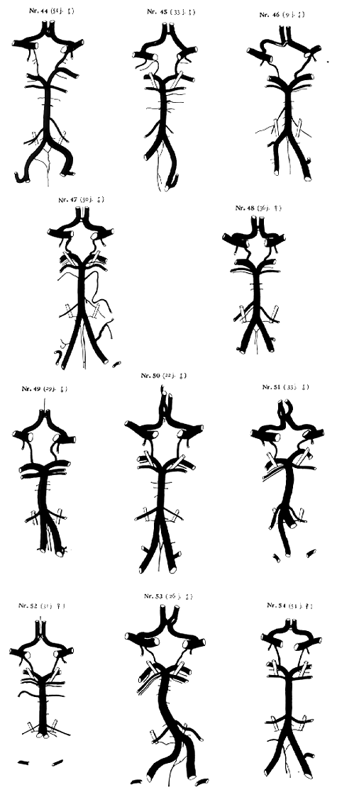
Anatomy Atlases. Immagine ospitata dal sito d’origine. Apri la pagina originale.
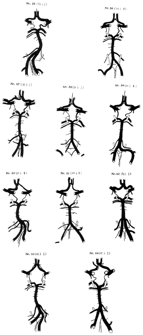
Anatomy Atlases. Immagine ospitata dal sito d’origine. Apri la pagina originale.
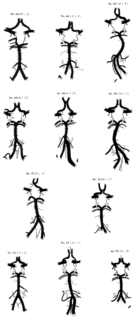
Anatomy Atlases. Immagine ospitata dal sito d’origine. Apri la pagina originale.
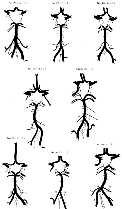
Anatomy Atlases. Immagine ospitata dal sito d’origine. Apri la pagina originale.
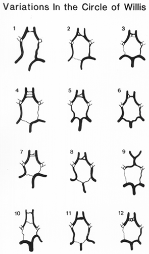
Anatomy Atlases. Immagine ospitata dal sito d’origine. Apri la pagina originale.
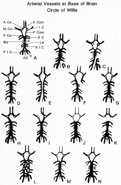
Anatomy Atlases. Immagine ospitata dal sito d’origine. Apri la pagina originale.
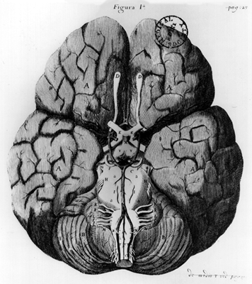
Anatomy Atlases. Immagine ospitata dal sito d’origine. Apri la pagina originale.
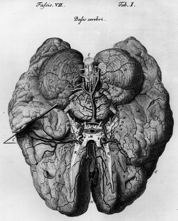
Anatomy Atlases. Immagine ospitata dal sito d’origine. Apri la pagina originale.
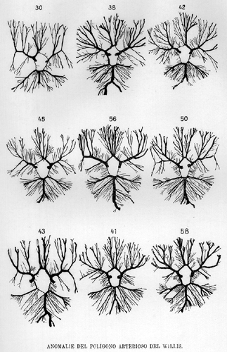
Anatomy Atlases. Immagine ospitata dal sito d’origine. Apri la pagina originale.
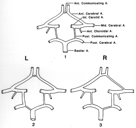
Anatomy Atlases. Immagine ospitata dal sito d’origine. Apri la pagina originale.
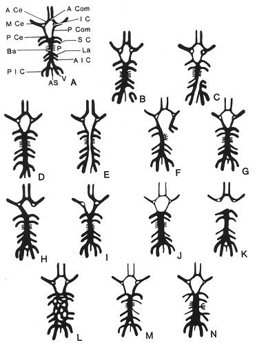
Anatomy Atlases. Immagine ospitata dal sito d’origine. Apri la pagina originale.
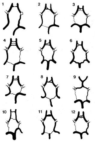
Anatomy Atlases. Immagine ospitata dal sito d’origine. Apri la pagina originale.
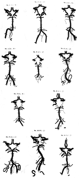
Anatomy Atlases. Immagine ospitata dal sito d’origine. Apri la pagina originale.
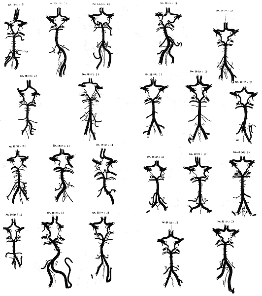
Anatomy Atlases. Immagine ospitata dal sito d’origine. Apri la pagina originale.
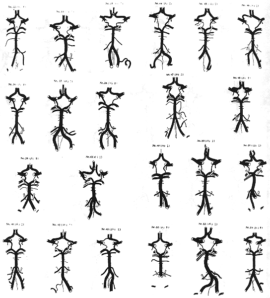
Anatomy Atlases. Immagine ospitata dal sito d’origine. Apri la pagina originale.
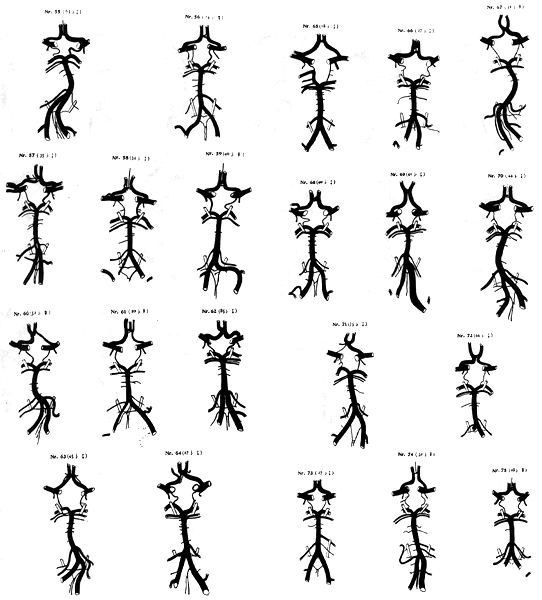
Anatomy Atlases. Immagine ospitata dal sito d’origine. Apri la pagina originale.
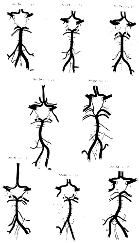
Anatomy Atlases. Immagine ospitata dal sito d’origine. Apri la pagina originale.
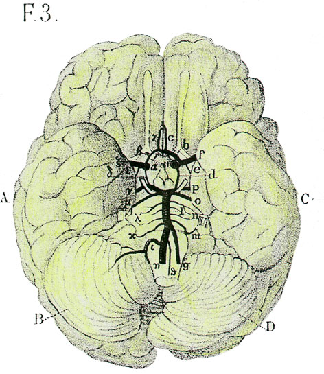
Anatomy Atlases. Immagine ospitata dal sito d’origine. Apri la pagina originale.
Riferimenti dell’opera originale
Elenco riportato come compare nella fonte, senza normalizzazione dei formati bibliografici.
- Baptista, A.G. (1964) Studies on the arteries of the brain. III. Circle of Willis: Morphologic features. Acta Neurol. Scand. 40:398-414.
- Beaumanoir, -. (1882) Anomalies des arteries de la base de l'encephale. Bull. et Mem. de la Societe Anatomique de Paris. 1882:317.
- Beevor, C.E. (1908) Cerebral blood supply Brain 30:403-425.
- Bergman, R.A., Thompson, S.A., Afifi, A.K. and F.A. Saadeh. (1988) Compendium of Human Anatomic Variation: Catalog, Atlas and World Literature. Urban & Schwarzenberg, Baltimore and Munich.
- Blackburn, J.W. (1907) Anomalies of encephalic arteries among the insane. A study of the arteries at the base of the encephalon in two hundred and twenty consecutive cases of mental disease, with special reference to anomalies of the circle of Willis. J. Comp. Neurol. 17:493-509.
- Calori, L. (1880) Di una inversione splancnica generale nell'uomo accompagnata da notabili del capo con esso lei convenienti e da estranee anomalie. Mem. R. Accad. Sci. Istituto di Bologna S. 4., 2:597-622.
- Cavatorti, P. (1907) Di una rara variazione delle arterie della base dell'encefalo nell'uomo. Monitore Zoologico Italiano 18:294-297.
- Chrzanowska, G. and J. Mierzwa. (1979) Variation of the cerebral arterial circle (of Willis) in man. Folia Morphol., Warsaw 38 (3):365-373.
- Decker, K. and E. Hipp (1958) Der basale Gefässkranz. Morphologie und Angiographie. Anat. Anz. 105:100-116.
- de Vriese, B. (1904) Sur les artères de la base du cerveau. Anatomischen Gesellschaft 1904: 88-99.
- de Vriese, B. (1904) Sur signification morphologique des artéries cérébrales. Arch. Biol. 21:357-457.
- Duroux, P.E., Dujul, H., Avet, J. and C. Gabrielle. (1953) A propos du polygone de Willis. Assoc. Anatomistes, Comptes Rendus. 40:328-338.
- Fawcett, E. and J.V. Blachford. (1906) The circle of Willis: an examination of 700 specimens. J. Anat. Physiol. 40:63-70.
- Garcia, D.F. (1895) Anomalias Arteriales. Revista de Ciencias Medicas 10:75-77.
- Grttler, K. (1951) Die bedeutung der funktionellen Struktur der Gefässwand. Gegenbaurs Morphol. Jahrbuch 91:368-393.
- Grttler, K. (1953) Die funktionelle Bedeutung des Baues der Gefasswand. Deutsche Z. Nervenheilkunde 170:433-445.
- Goldstein, M. (1914) Beitrag zur Anatomie und funktionellen Bedeutung der Arterien des Gehirns, inbesondere des Balkens. Z. Ges. Neurol. Psychiatr. 26:361-396.
- Grenell, R.G. (1953) Central Nervesystem resistance. IV Age and resistance to temporary arrest of the cerebral circulation. J. Comp. Neurol. 99:117-134.
- Gunter, R.G. (1964) The diameter of the cerebral arteries of the human fetus. Anat. Rec. 150:79-88.
- Hasenjäger, T. (1937) Ein Beitrag zu den Abnormitäten des Circulus Wilisii. Zbl. Neurochir. 2:34- 38.
- Kaplan, H.A. and D.H.Ford. (1966) The Brain Vascular System. Elsevier, Amsterdam.
- Kirgis, J.D., Fisher, W.L., Llewellyn, R.C. and E. McC. Peebles. (1966) Aneurysms of anterior communicating artery and gross anomalies of the circle of Willis. J. Neurosurg. 25:73-78.
- Kleiss, E. (1941-42) Die verschiedenen Formen des Circulus arteriosus Willisi. Anat. Anz. 92:216-230.
- Kmentowa-Sklenska, A. and Z. Hejlowa. (1958) Struktura mpzgovych tepen u dospivajiciho a dospeleho cloveka. Ceskoslov. Morph. 6:358-367.
- Kohman, St. (1957) Uklad tetnic podstawy mozgowia czlowieka. Folia Morphol., Warsaw 8:229-239.
- Latarjet, A. (1948) Testut's Traite d'Anatomie Humaine, 9th ed., G. Doin & Cie., Paris.
- LeGuyader, A., Cayotte, J.L., Laffont, J., Renard, M., Delteil, C., Picard, L., Brice, M. and J.P. Masson. (1968) Variations morphologiques du polygone de Willis à propos de 170 observations anatomiques. Étud. Anat. Fac. Med., Nancy 2:45-55.
- Longo, L. (1905) Le anomalie del poligono di Willis nell'uomo studiate comparativamente in alcuni mammiferi ed ucelli. Anat. Anz. 27:170-176.
- Lowrey, L.G. (1916) Anomaly in the circle of Willis, due to absence of the right internal carotid artery. Anat. Rec. 10:221-222.
- McCullough, A.W. (1962) Some anomalies of the cerebral arterial circle (of Willis) and related vessels. Anat. Rec. 142:537-543.
- McDonald, D. A. and J.M. Potter. (1951) The distribution of the blood to the brain. J. Physiol., London 114:356-371.
- McMinn, R.M.H. (1953) A case of non-union of the vertebral arteries. Anat. Rec. 116:283-286.
- Miller-Fisher, C. (1965) The circle of Willis. Anatomical variations. Vasc. Dis. 2:99-105.
- Mittenwallner, F. (1955) A Variationsstatistische Untersuchungen an den basalen Hirngefässen. Acta Anat., Basel 24:51-88.
- Mori, A. (1898) Sulla disposizione delle arterie della base dell'encefalo nei normali e negli alienati. Monitore Zoologico Italiano 4:179-202.
- Opitz, E. and M. Schneider. (1950) über die Sauerstoffversogung des Gehirns und dem Mechanimus von Mangelwirkung. Ergeb. Physiol.. Biol. Chemie., Experim. Pharmak. 46:116-126.
- Parnisetti, C. (1902) Anomalie del poligono arterioso del Willis nei delinquenti in rapporto con alterazioni del cervello e del cuore. Archivio di Psichiatria, Scienze Penali ed Antropologia Criminale (vol. 7 della Serie 2)23:11-27.
- Poynter, C.W.M. (1922) Congenital anomalies of the arteries and veins of the human body with bibliography. The University Studies of the University of Nebraska, Lincoln 22:1-106.
- Puchades-Orts, A., Nombela-Gomez, M. and G. Ortuno-Pacheco. (1976) Variations in form of circle of Willis: some anatomical and embryological considerations. Anat. Rec. 185(l):119-123.
- Riggs, H. and C.Rupp. (1963) Variations in form of the circle of Willis. The relation of the variations to collateral circulation, anatomic analysis. Arch. Neurol. 8:24-30.
- Slany, A. (1938) Anomalien des Circulus arteriosus Willisi in ihrer Beziehung zur Aneurysmenbildung an der Hirnbasis. Arch. Pathol. Anat. Physiol. Klin. Med. 301:62-71.
- Stebbens, W.E. (1963) Aneurysms and anatomical variation of cerebral vessels. Arch. Pathol. 75:45-64.
- Stopford, J.S.B. (1916) The arteries of the pons and medulla oblongata. J. Anat. Physiol. 50:131-164.
- Waddington, M.M. (1974) Atlas of Cerebral Angiography with Anatomic Correlation. Little, Brown and Co., Boston.
- Willis, T. (1664) Cerebri anatomie: cui accessit nervorum descripto et usus. Martyn and Allestry. London.
- Windle, B.C.A. (1888) On the circle of Willis. New York Medical Journal 2:XXX-XXX.
- Windle, B.C.A. (1888) The arteries forming the circle of Willis. J. Anat. Physiol. 22:289-293.
Classificazione anatomica
- Sistemi:
- Sistema cardiovascolare
Provenienza del contenuto
Adattamento
Adattamento di «Circle of Willis — Illustrated Encyclopedia of Human Anatomic Variation: Opus I: Muscular System» di Ronald A. Bergman, Adel K. Afifi, Ryosuke Miyauchi. Fonte: https://www.anatomyatlases.org/AnatomicVariants/Cardiovascular/Text/Arteries/CircleofWillis.shtml Opera originale distribuita con licenza CC BY-NC-SA 4.0 (https://creativecommons.org/licenses/by-nc-sa/4.0/deed.it). Adattamento a cura di Redazione AIVA. Modifiche rispetto all’originale: Testo originale inglese riprodotto integralmente e riorganizzato nel formato scheda del portale (sintesi, testo, riferimenti), senza modificarne le affermazioni. La traduzione italiana non è ancora stata eseguita, e la pagina lo dichiara. Le figure non sono state copiate: sono collegate alla pagina d’origine. Questa pubblicazione non è approvata né avallata dagli autori dell’opera originale.
Licenza
Questo contenuto è distribuito con licenza CC BY-NC-SA 4.0. Licenze e attribuzioni