Voce enciclopedica
Common Interosseous Artery
Abstract
The palmar and dorsal (posterior) vessels may arise separately from the ulnar. A high origin of the common interosseous has been described as arising from the axillary artery. Palmar interosseous arteries may be so well-developed that they substitute for branches of the ulnar and/or radial arteries The common interosseous may be a branch of a high radial artery and it may provide the radial artery deep to pronator teres. A usual anterior interosseous may be absent and be replaced by branches of the radial artery.
The palmar and dorsal (posterior) vessels may arise separately from the ulnar.
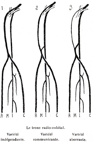
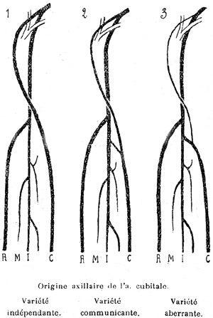
A high origin of the common interosseous has been described as arising from the axillary artery.
Palmar interosseous arteries may be so well-developed that they substitute for branches of the ulnar and/or radial arteries
The common interosseous may be a branch of a high radial artery and it may provide the radial artery deep to pronator teres.
A usual anterior interosseous may be absent and be replaced by branches of the radial artery.
It may join the radial artery at the wrist, or if the radial stops short of the carpus the posterior interosseous artery may replace it.
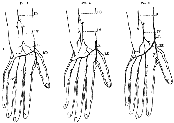
One of the branches of the anterior interosseous is of special interest. The median artery is a long slender artery that arises from the proximal part of the interosseous and supplies the median nerve throughout its course in the forearm. Occasionally (~8%), the median arterial branch is large and continues into the hand, deep to the flexor retinaculum along with the median nerve to join in the formation of the superficial palmar arch. The addition of the median artery to the already numerous and crowded carpal canal components may compress the median nerve and lead to the carpal tunnel syndrome.
The median artery is a persistence of the developmental pattern, in which the median artery maintains the superficial palmar network while the radial and ulnar arteries are developing. It persists as a variant, even after the final form of the development of the radial and ulnar vessels in the hand has occurred. The median artery usually arises from the common interosseous artery or the anterior interosseous.
The recurrent interosseous branch of the posterior interosseous is frequently missing and may be replaced by a branch from the common interosseous artery. The posterior interosseous may give rise to an accessory posterior interosseous; it may also give rise to a superficial external interosseous artery that courses superficial to flexor carpi ulnaris to the wrist. High Origin of the Arteries of the Forearm Radio-Ulnar-Interosseous Trunk
Number of Observations Total Origin:
1.
0
2.
270 24 ?
3.
136 19 ?
1) Statistics by Giacomini. 2) Statistics by authors cited by Giacomini. 3) Statistics by Dubreuil-Chambardel.
Anterior
Anatomy Atlases. Immagine ospitata dal sito d’origine. Apri la pagina originale.
Anterior
Anatomy Atlases. Immagine ospitata dal sito d’origine. Apri la pagina originale.
Anterior
Anatomy Atlases. Immagine ospitata dal sito d’origine. Apri la pagina originale.
Anterior
Anatomy Atlases. Immagine ospitata dal sito d’origine. Apri la pagina originale.
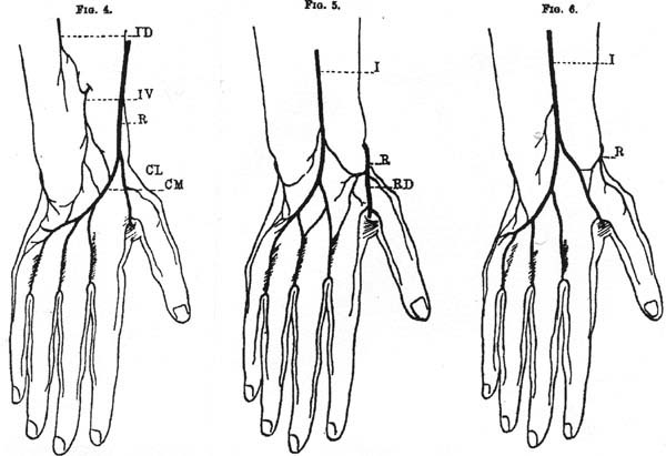
Anterior
Anatomy Atlases. Immagine ospitata dal sito d’origine. Apri la pagina originale.
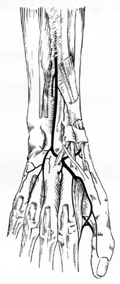
Posterior
Anatomy Atlases. Immagine ospitata dal sito d’origine. Apri la pagina originale.
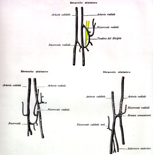
Posterior
Anatomy Atlases. Immagine ospitata dal sito d’origine. Apri la pagina originale.
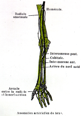
Posterior
Anatomy Atlases. Immagine ospitata dal sito d’origine. Apri la pagina originale.
See Image 250
Superficial Median
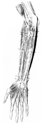
Anatomy Atlases. Immagine ospitata dal sito d’origine. Apri la pagina originale.
Riferimenti dell’opera originale
Elenco riportato come compare nella fonte, senza normalizzazione dei formati bibliografici.
- Anson, B.J., Ed. (1966) Morris' Human Anatomy, 12th ed., The Blakiston Division, McGraw-Hill Book Company, New York.
- Bergman, R.A., Thompson, S.A., Afifi, A.K. and F.A. Saadeh. (1988) Compendium of Human Anatomic Variation: Catalog, Atlas and World Literature. Urban & Schwarzenberg, Baltimore and Munich.
- Broca, -. (1849) Anomalies artérielles d'un member thoracique. Perforation du nerf median par l'artère inter osseuse. Bulletins et Mem. de la Société Anatomique de Paris XXIV(3):67-70.
- Henle, J. (1868) Handbuch der Systematischen Anatomie des Menschen. 3 vols. Von Friedrich Vieweg und Sohn., Braunschweig.
- Latarjet, A. (1948) Testut's Traite d'Anatomie Humaine, 9th ed., G.Doin & Cie., Paris.
- Parmentier, -. (1850) Anomalies artérielles; un cas d'origine prématuree de l'artère interosseuse. Bulls. et Méms. de la Soc. Anatomique de Paris 1850:75.
- Salvi, G. (1898) Arteriae dorsalis carpi. Atti della Societia a Toscana di Scienze Naturali, residente in Pisa. 17:167-185.
- Schaefer, E.A., Symington, J, and T.H. Bryce, Eds. Quain's Anatomy, 11th ed., Longmans, Green and Co., London.
- Tandler, J. (1897) über die Varietäten der Arterien der Hand. Anat. Hefte 7:263-282.
Classificazione anatomica
- Sistemi:
- Sistema cardiovascolare
Provenienza del contenuto
Adattamento
Adattamento di «Common Interosseous Artery — Illustrated Encyclopedia of Human Anatomic Variation: Opus I: Muscular System» di Ronald A. Bergman, Adel K. Afifi, Ryosuke Miyauchi. Fonte: https://www.anatomyatlases.org/AnatomicVariants/Cardiovascular/Text/Arteries/InterosseousCommon.shtml Opera originale distribuita con licenza CC BY-NC-SA 4.0 (https://creativecommons.org/licenses/by-nc-sa/4.0/deed.it). Adattamento a cura di Redazione AIVA. Modifiche rispetto all’originale: Testo originale inglese riprodotto integralmente e riorganizzato nel formato scheda del portale (sintesi, testo, riferimenti), senza modificarne le affermazioni. La traduzione italiana non è ancora stata eseguita, e la pagina lo dichiara. Le figure non sono state copiate: sono collegate alla pagina d’origine. Questa pubblicazione non è approvata né avallata dagli autori dell’opera originale.
Licenza
Questo contenuto è distribuito con licenza CC BY-NC-SA 4.0. Licenze e attribuzioni