Voce enciclopedica
Femore e rotula
Abstract
Il femore può mancare, in tutto o nel solo corpo, e presentare un terzo trocantere, una fossa ipotrocanterica o un processo sopracondiloideo. La rotula è talvolta bipartita e ne è stata riportata l’assenza congenita.
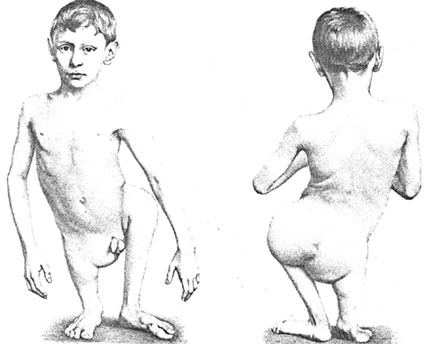
È stata riportata l’assenza congenita bilaterale del corpo del femore ed è stata descritta anche l’assenza unilaterale dell’intero femore.
Occasionalmente la tuberosità glutea è considerevolmente ingrandita e viene denominata terzo trocantere. La tuberosità glutea può anche essere assente e, al suo posto, l’area può presentare una fossa ipotrocanterica. In alcuni casi la tuberosità e la fossa coesistono.
Anche lo sviluppo della linea aspra può mostrare una variazione considerevole.
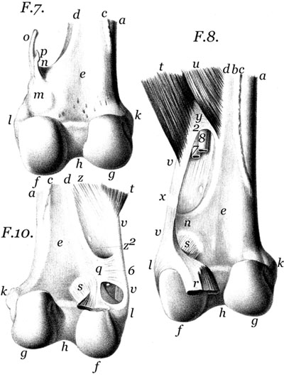
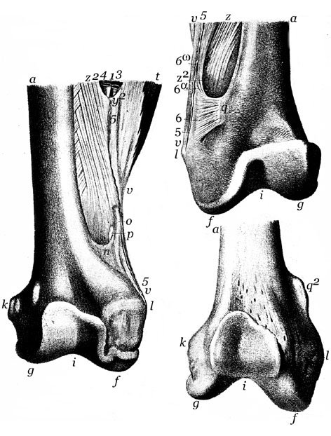
È stato descritto un processo osseo, lungo circa 3 cm, che origina in corrispondenza dell’origine del capo breve del bicipite femorale. Da alcuni è ritenuto analogo al processo sopracondiloideo dell’omero.
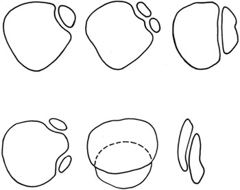
La rotula è talvolta bipartita, probabilmente per la mancata unione dei centri di ossificazione, e l’angolo prossimale laterale è quello che più frequentemente si riscontra come frammento separato. È stata riportata l’assenza congenita della rotula. Nei casi di assenza congenita del muscolo quadricipite la rotula può essere presente, ma è di solito lussata.
Assenza congenita dei femori.
Anatomy Atlases. Immagine ospitata dal sito d’origine. Apri la pagina originale.
Processo sopracondiloideo mediale del femore.
Anatomy Atlases. Immagine ospitata dal sito d’origine. Apri la pagina originale.
Processo sopracondiloideo mediale del femore.
Anatomy Atlases. Immagine ospitata dal sito d’origine. Apri la pagina originale.
Variazioni delle forme di patella partita.
Anatomy Atlases. Immagine ospitata dal sito d’origine. Apri la pagina originale.
Riferimenti dell’opera originale
Elenco riportato come compare nella fonte, senza normalizzazione dei formati bibliografici. Le voci non sono tradotte: un riferimento bibliografico si cita nella lingua in cui è pubblicato.
- Adams, J.D. and D.R. Leonard. (1925) A development anomaly of the patella frequently diagnosed as fracture. Surg., Gynecol. Obstet. 41:601-604.
- Brücki, H. von. (1942) Über die Patella cubiti, eine seltene Abweichung des Ellenbogenskelettes. Z. Orthop. 73:158-160.
- Brunner, G. (1891) Über Genese, congenitalen Mangel und rudimentäre Bildung der Patella. Arch. Pathol. Anat. Physiol. Klin. Med. 124:358-373.
- Burguières, -. (1837) Absence congéniale du col du femur. Bulletins et Memoires de la Société Anatomique de Paris XII(8)(1837):226-227.
- Dide, M. (18 93) Note sur une apophyse anormale recontrée sur un fémur humain. Bull. de la Soc. d'Anthrop. de Paris 4:162-165.
- Duhem, -. (1925) Malformations osseuses multiple avec fémur bifide. Bull. et Mém. de la Soc. d'Anthrop. de Paris 6:9-10.
- Frantz, C.H. and R. O'Rahilly. (1961) Congenital skeletal limb deficencies. J. Bone Joint Surg. (Am.) 43:1202-1224.
- Gasco, J., Del Pino, J.M. and F. Gomar-Sancho. (1987) Double patella: A case of duplication in the coronal plane. J. Bone Joint Surg. (Brit) 69:602-603.
- Giles, R.G. (1928) A congenital anomaly of the patella. Texas State J. Med. 23:731-732.
- Grashey, R. (1937) Dorsale supracondyläre Femurexostose. Roentgenpraxis 9:278-280.
- Greeley, P.W. (1932) Multiple ossification centers of the patella. Am. J. Surg. 18:456-459.
- Gruber, W. (1883) In Bildungsanomalien mit Bildungshemmung begründete Bipartition beider Patellae eines jungen Subjects. Arch. Pathol. Anat. Physiol. Klin Med. 94:358-361.
- Henkel, L. and H.G. Willert. (1969) Dymelia. A classification and a pattern of malformation in a group of congenital defects of the limbs. J. Bone Joint Surg. (Br.) 51:399-414.
- Hyrtl, -. (1858) Ein Fall von Processus supracondyloidens femoris (Gruber) an Lebenden. Sitzungsberichte der Wiener Akad. der Wissen. Math. -Natur. Kl. 31:231-234.
- Jones, H.C. and D.W. Hedrick. (1942) Patellar anomalies, roentgenologic and clinical consideration. Radiology 38:30-34.
- Jovanin, T., Dupont, J.Y., Halimi, P. and J.P. Lassau. (1982) The synovial folds of the knee joint: An anatomical study. Anat. Clin. 4:47-53.
- Katdare, S.S. (1945) Congenital absence of the shaft of the femur on both sides. Indian Med. Gaz. 80:297-298.
- Kate, B.R. (1964) A study of the regional variation of the Indian femur - the diameter of the head - its medico legal and surgical application. J. Anat. Soc. India 13:80-84.
- Kempson, F.C. (1902) Emargination of the patella. J. Anat. Physiol. 36:419-420.
- Kiil-Nielsen, K. (1944) Case of congenital absence of the patella and its treatment. Acta Orthop. Scandianav. 15:49-57.
- Kutz, E.R. (1949). Congenital absence of the patellae. J. Pediatr. 34:760-762.
- Langston, H.H. (1939-40) Congenital defect of the shaft of the femur. Brit. J. Surg. 27:162-165.
- Manohar, K. (1939-40) Congenital absence of the right femur. Brit. J. Surg. 27:158-161.
- Manouvrier, L. (1893) Etude sur les variations morphologiques du corps du fémur dans lespèce humaine. Bull. de la Soc. d'Anthrop. de Paris 4:111-144.
- Nadgir, Y. G. (1917) The supracondyloid tubercles of the femur. J. Anat. 51:375.
- Oetteking, B. (1923) Anomalous patellae. Anat. Rec. 23:269-279.
- Pires de Lima, J.A. (1913) On a bone found in the musculus vastus externus of the thigh. J. Anat. Physiol. 47:414-424.
- Pittard, E., Manula, L. and A. Manula. (1946) Troisème trochanter et fosse hypotrochantérienne chez 500 fémurs humains (250 de chaque sexe) provenant de la région de Genève. Caractères fonctionnels ou caractéres réversifs? Actes Soc. Helvétiques Sci. Nat. 1946:175-176. Cited in Excerpta Medica, Sec. 1, Vol. 2, abstract 1372, p. 484, 1948.
- Roudenko, M.S. (1914) Note sur les variations de l'inclinasion de la tête du fémur et sur la hauteur ischio-suscotyloideinne suivant l'indice bustoskelique. Bull. et Mém. de la Soc. d'Anthropol. de Paris 5:200-204.
- Senturia, H.R. and B.D. Senturia. (1944) Congenital absence of the patellae associated with arthrodysplasia of the elbows and dystrophy of the nails. A hereditary syndrome. Am. J. Roentgenol. 51:352-358.
- Shah, M.A. (1945) The popliteal facet in men who habitually squat. Nature 156:237-238.
- Siddiqi, M.A.H. (1933-34) Variations in the lower end of the femur from Indians. J. Anat. 68:331-337.
- Siddiqi, M.A.H. ((1935-36) Observations on the lower end of the femur from American Indians and modern Canadians. J. Anat. 70:410-412.
- De Souza Fontes, J.R. (1877) Ein Lebender Knabe mit angeborenem Mangel der Glieder. Arch. Pathol. Anat. Physiol. Klin. Med. 71:107-111.
- Stoppord, J.S.B. (1915) The supracondyloid tubercles of the femur and the attachment of the gastronemius muscle to the femoral diaphysis. J. Anat. Physiol. 49:80-84.
- Sunderland, S. (1938) The quadrate tubercle of the femur. J. Anat. 72:309-312.
- Susman, M.P. (1934) Rudimentary patellae, other skeletal defects, and dystrophy of the nails. Med. J. Australia 1:685-686.
- Swaton, S. and Z. Huber. (1960) Einseitige, angeborene, doppelte Kniescheibe (Patella duplex unilateralis congenital). Zentralbl. Chir. 85:2270-2274.
- Todd, T.W. and W.C. McCally. (1921) Defects of the patellar border. Ann. Surg. 74:775-782.
- Treves, F. (1887) A gluteal trochanter in the human subject. J. Anat. Physiol. 21:325-327.
- Williams, W.R. (1884) Congenital absence of the femora. Trans. Path. Soc. Lond. 35:317-318.
- Wright, W. (1904) Case of accessory patellae in the human subject, with remarks on emargination of the patella. J. Anat. Physiol. 38:65-67.
- Wütschke, J. (1953) Ein Fall von linksseitiger doppeler Patella. Fortschr. Röntgenstr. 78:218-220.
- Wütschke, (1966) J. Patella partita und Patella duplex (kritische Betrachtung zur Differentialdiagnose). Fortschr. Roentgenstr. 104:260-263.
Classificazione anatomica
- Sistemi:
- Sistema scheletrico
Provenienza del contenuto
Traduzione
Traduzione italiana di «Femur and Patella — Illustrated Encyclopedia of Human Anatomic Variation: Opus I: Muscular System» di Ronald A. Bergman, Adel K. Afifi, Ryosuke Miyauchi. Fonte: https://www.anatomyatlases.org/AnatomicVariants/SkeletalSystem/Text/FemurPatella.shtml Opera originale distribuita con licenza CC BY-NC-SA 4.0 (https://creativecommons.org/licenses/by-nc-sa/4.0/deed.it). Traduzione a cura di Redazione AIVA. Modifiche rispetto all’originale: Traduzione italiana del testo originale inglese, che resta consultabile nella versione inglese di questa pagina. Il contenuto è stato riorganizzato nel formato scheda del portale (sintesi, testo, riferimenti) senza modificarne le affermazioni. Le figure non sono state copiate: sono collegate alla pagina d’origine. Questa pubblicazione non è approvata né avallata dagli autori dell’opera originale.
Licenza
Questo contenuto è distribuito con licenza CC BY-NC-SA 4.0. Licenze e attribuzioni
