Voce enciclopedica
Iliopsoas (Hyrtl) (Iliacus and Psoas)
Abstract
The psoas and iliacus muscles are occasionally completely independent muscles. The psoas may be divided longitudinally into fascicles. An accessory slip has been described lateral to the muscle and separated from it by the femoral nerve. Iliacus may be pierced by the femoral nerve. Mori reports that the psoas major arose from the first lumbar vertebra in 98.6 % of his 152 sides (76 cadavers), and from the second lumbar on two sides, 1.3%. In no instance did the muscle arise from thoracic vertebrae.
The psoas and iliacus muscles are occasionally completely independent muscles.
The psoas may be divided longitudinally into fascicles. An accessory slip has been described lateral to the muscle and separated from it by the femoral nerve. Iliacus may be pierced by the femoral nerve.
Mori reports that the psoas major arose from the first lumbar vertebra in 98.6 % of his 152 sides (76 cadavers), and from the second lumbar on two sides, 1.3%. In no instance did the muscle arise from thoracic vertebrae.
Macalister published the following:
- The two or three lower vertebral origins, or the upper oblique-process-origin of the medial head, may give a slip separate from the remainder of the muscle (Albinus, Meckel). This may receive a slip from the psoas parvus (minor) tendon
- A bundle of the lateral head may leap over one or more roots of the crural (femoral) nerve
- A second or lesser psoas (minor), lying between psoas magnus (major) and iliacus, and going to the lesser trochanter, is described by Meckel. Horner (Philadelphia) has seen several slips forming psoas major, and the same author has seen this muscle perfectly separated from iliacus, an anomaly of very rare occurrence, as they are nearly always inserted at least by a common tendon. Macalister and Lieutaud both have seen the same division
- Macalister has traced a band of fibers of the psoas, springing from the ligamentum arcuatum internum, (medial arcuate ligament of diaphragm), and one continuous with those of the right crus
- The psoas major may have an origin from the last lumbar vertebra, but most commonly is either not at all, or slightly connected to that bone.
In regard to iliacus, Macalister reported the following:
- Iliacus may be found divided into two parts, or with a partly separated superficial lamina arising from the fascia iliaca, or with a slip from the psoas crossing over the crural (femoral) nerve, and become continuous with it
- A separate slip occasionally arises from the ilio-pectineal line, and sometimes another from the inner margin of the incisura semilunaris of the ilium
- A few fibers from the iliolumbar part of quadratus lumborum are continuousl occasionally into the origin of iliacus and its most posterior fibers, which frequuently are attached to the iliosacral ligaments, may extend to the promontory of the sacrum.
Iliacus minor or iliocapsularis (third head of iliacus), a small detached portion of iliacus, is frequently present. It arises from the anterior inferior spine of the iliac bone and inserts onto the lower part of the intertrochanteric line of the femur, or into the iliofemoral ligament. It is usually separated from iliacus by arising either direclty from the margin, or from its outer surface. Its insertion is always above the main iliacus tendon.
A slip from iliacus may run medial to the psoas muscle.
Syn.: m. Iliacopsoas (Haller), Flexor Femoris (Theile), Psoas-Iliaque (Cruveilheir), Psoas Magnus (Macalister).
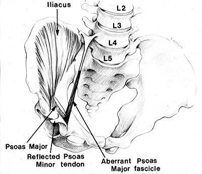
Psoas Major Accessorius.
from Saadeh and Bergman — Anatomy Atlases. Immagine ospitata dal sito d’origine. Apri la pagina originale.
Riferimenti dell’opera originale
Elenco riportato come compare nella fonte, senza normalizzazione dei formati bibliografici.
- Anson, B.J., Ed. (1966) Morris' Human Anatomy,12th ed., The Blakiston Division, McGraw-Hill Book Company, New York.
- Bonin, G. von. (1930) Bau und Variationen des Iliopsoas. Anat. Anz. 69:476-485.
- Clarkson, R.D. and H. Rainy. (1889) Unusual arrangement of the psoas muscle. J. Anat. Physiol. 23:504-506.
- Macalister, A. (1875) Observations on muscular anomalies in the human anatomy. Third series with a catalogue of the principal muscular variations hitherto published. Trans. Roy. Irish Acad. Sci. 25:1-130.
- Mori, M. (1964) Statistics on the musculature of the Japanese. Okajimas Fol. Anat. Jap. 40: 195-300.
- Saadeh, F.A. and R.A. Bergman. (1985) An aberrant psoas major muscle fascicle. Anat. Anz. 160:367-368.
- Schaefer, E.A., Symington, J., and T.H. Bryce., Eds. (1923) Quain's Anatomy, 11th ed., Longmans Green and Co., London.
Classificazione anatomica
- Sistemi:
- Sistema muscolare
Provenienza del contenuto
Adattamento
Adattamento di «Iliopsoas (Hyrtl) (Iliacus and Psoas) — Illustrated Encyclopedia of Human Anatomic Variation: Opus I: Muscular System» di Ronald A. Bergman, Adel K. Afifi, Ryosuke Miyauchi. Fonte: https://www.anatomyatlases.org/AnatomicVariants/MuscularSystem/Text/I/06Iliopsoas.shtml Opera originale distribuita con licenza CC BY-NC-SA 4.0 (https://creativecommons.org/licenses/by-nc-sa/4.0/deed.it). Adattamento a cura di Redazione AIVA. Modifiche rispetto all’originale: Testo originale inglese riprodotto integralmente e riorganizzato nel formato scheda del portale (sintesi, testo, riferimenti), senza modificarne le affermazioni. La traduzione italiana non è ancora stata eseguita, e la pagina lo dichiara. Le figure non sono state copiate: sono collegate alla pagina d’origine. Questa pubblicazione non è approvata né avallata dagli autori dell’opera originale.
Licenza
Questo contenuto è distribuito con licenza CC BY-NC-SA 4.0. Licenze e attribuzioni