Voce enciclopedica
Knee
Abstract
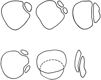
In a study of 433 knee joints, the menisci were found to be "C"-shaped in 422, discoid in nine, and circular in two. The medial meniscus may not be "C"-shaped but rather a ring completed by a ligament. In 140 knees, a menicofemoral ligament was present in 99 (71%). In 50 cases (36%) the ligament was the anterior meniscofemoral and in the remaining 49 cases (35%) it was the posterior meniscofemoral ligament. In 17 cases (12%) the anterior ligament was bilateral, in 12 cases (8%) the posterior was bilateral, and in two cadavers both ligaments were found in both knees.
In a study of 433 knee joints, the menisci were found to be "C"-shaped in 422, discoid in nine, and circular in two. The medial meniscus may not be "C"-shaped but rather a ring completed by a ligament.
In 140 knees, a menicofemoral ligament was present in 99 (71%). In 50 cases (36%) the ligament was the anterior meniscofemoral and in the remaining 49 cases (35%) it was the posterior meniscofemoral ligament. In 17 cases (12%) the anterior ligament was bilateral, in 12 cases (8%) the posterior was bilateral, and in two cadavers both ligaments were found in both knees. No sex differences were found.
The cruciate ligaments may have accessory slips or appendices. The anterior cruciate ligament may be absent.
The posterior horn of the medial meniscus occasionally supports a free-lying cartilage; which may be anchored posteriorly to the tibia. A lateral interarticular disk may be present.
The transverse ligament may be variably developed or absent.
The patella may vary in size and position. It may exist in several parts or it may be absent!
See also Thigh: Femur and Patella .
Anatomy Atlases. Immagine ospitata dal sito d’origine. Apri la pagina originale.
Riferimenti dell’opera originale
Elenco riportato come compare nella fonte, senza normalizzazione dei formati bibliografici.
- Barucha, E. (1960) Unsere Erfahrungen über den Wert des Rauberschen Röntgen-Zeichens bei der Meniscus-Diagnose. Monatsschr. Unfallheilk. 63:370-375.
- Basmajian, J.V. (1952) A ring shaped medial semi-lunar cartilage. J. Bone Joint Surg. (Br.) 34:638-639.
- Charles, C.M. (1935) On the menisci of the knee joint in American whites and negroes. Anat. Rec. 63:355-364.
- Gruber, W. (1883) In Bildungsanomalie mit begründete Bipartition beider Patellae eines jungen Subject. Arch. f. Pathol Anat. Physiol. Klin. Med., Berlin 94:353-357.
- Heller, L. and J. Langman. (1964) The menisco-femoral ligaments of the human knee. J. Bone Joint Surg. (Br.) 46:307-313.
- Kaplan, E.B. (1957) Discoid lateral meniscus of the knee joint. J. Bone Joint Surg. (Am.) 39:77-87.
- Kiil-Nielsen, Kr. (1944) Case of congenital absence of the patella and its treatment. Acta Orthop. Scandinav. 15:49-57.
- Richmond, D.A. (1958) Two cases of discoid medial cartilage. J. Bone Joint Surg. (Br.) 40:268-269.
- Ross, J.A., Tough, I.C.K. and T.A. English. (1958) Congenital discoid cartilage. Report of a case of discoid medial cartilage, with an embryological note. J. Bone Joint Surg. (Br.) 40:262-267.
- Vermes, E. (1923) Ein Fall von hochgrädiger Anomalie im inneren Bandapparat eines Kniegelenkes. Anat. Anz. 56:427-430.
- Vermes, E. (1923-24) Ein Beitrag zu den Varietäten der Bänder im Inneren des Kniegelenkes. Anat. Anz. 57:218-221.
- Wuth, E.A. (1899) über angeborenen Mangel, sowie Herkunft und Zweck der Kniescheibe. Arch. Klin. Chir. 58:900-917.
- Wutzer, C.W. (1835) Angeborene Missbildungen des Kniegelenks. Arch. Anat. Physiol. 1835:385-390.
Classificazione anatomica
- Sistemi:
- Sistema scheletrico
Provenienza del contenuto
Adattamento
Adattamento di «Knee — Illustrated Encyclopedia of Human Anatomic Variation: Opus I: Muscular System» di Ronald A. Bergman, Adel K. Afifi, Ryosuke Miyauchi. Fonte: https://www.anatomyatlases.org/AnatomicVariants/SkeletalSystem/Text/Knee.shtml Opera originale distribuita con licenza CC BY-NC-SA 4.0 (https://creativecommons.org/licenses/by-nc-sa/4.0/deed.it). Adattamento a cura di Redazione AIVA. Modifiche rispetto all’originale: Testo originale inglese riprodotto integralmente e riorganizzato nel formato scheda del portale (sintesi, testo, riferimenti), senza modificarne le affermazioni. La traduzione italiana non è ancora stata eseguita, e la pagina lo dichiara. Le figure non sono state copiate: sono collegate alla pagina d’origine. Questa pubblicazione non è approvata né avallata dagli autori dell’opera originale.
Licenza
Questo contenuto è distribuito con licenza CC BY-NC-SA 4.0. Licenze e attribuzioni
