Voce enciclopedica
Latissimus Dorsi
Abstract
Variations in the extent of this muscle and its attachments are considerable. The muscle may arise only from the ribs rather from the vertebral column, ribs, scapula, and the crest of the ilium as is most common. The costal origin may be reduced to rib 12. Mori has reported the vertebral, costal, and scapular origins for latissimus dorsi from 50 Japanese cadavers, 100 halves, as follows: Vertebral Origin Number of Sides Upper Limit Right Left Total % T.
Variations in the extent of this muscle and its attachments are considerable. The muscle may arise only from the ribs rather from the vertebral column, ribs, scapula, and the crest of the ilium as is most common. The costal origin may be reduced to rib 12.
Mori has reported the vertebral, costal, and scapular origins for latissimus dorsi from 50 Japanese cadavers, 100 halves, as follows: Vertebral Origin
Number of Sides
Upper Limit
Right
Left
Total
%
T.
5
1
1
2
2
6
9
7
16
16
7
21
23
44
44
8
15
15
30
30
9
4
4
8
8 Costal Origin
Number of Sides
Rib
Right
Left
Total
%
9,10, 11,12
13
12
25
41.6
-,10,11,12
17
18
35
58.3 Scapular Origin
Bilateral
Unilateral
Right Side
Left Side
53.3%
8%
6%
The muscle may be divided into separate fascicles, frequently with one arising from the scapula. The muscle may be continuous with teres major.
Macalister described this muscle as "very liable to vary."
- It may have an origin from the ninth rib (Cloquet, Paxton)
- Or from the eight lower thoracic spines
- Or from the ninth intercostal fascia (Wood)
- The iliac origin may be suppressed
- Or reduced to a rudiment
- Macalister found it extending so far forward as to overlap the external oblique
- It frequently has a slip from the inferior margin of the scapula, as described by the majority of anatomists.
- Macalister has seen it rising no higher, in point of origin, than the first lumbar spine
- He has also seen the twelfth rib slip deficient, the muscle having but two costal orgins. Also seen by Sömmerring
- A slip extending from latissimus dorsi may cross the axillary space to the tendon of pectoralis major and has been named axillary arch, achselbogen, or pectorodorsalis muscle. The axillary arch may be fused with teres major. It may extend to the brachial fascia of coracobrachialis, to the tendon of the long head of the biceps, to the first rib, to the axillary fascia, to pectoralis minor, to pectoralis quartus, or to the coracoid process. Axillary arch muscles were discovered by Ramsay in 1795 and have a dissecting room frequency of 7%. Langer (1846) is also credited with a very early description of the axillary arch. See heading Axillary Arch . Its frequency is given as one in thirty by Meckel, eight in one hundred and five by Struthers, six in one hundred and six by Wood. All sources totaled it occurs once in sixteen subjects (6.25%). Struthers reported it was more common in females than males, but others do not find a sex link
- The achselbogen from latissimus dorsi may be attached to the lower border of pectoralis major
- Or to the fascia over biceps
- Or to the fascia over coracobrachialis
- In one singular case it coexisted with a second slip to the fascia over the fifth digitation of serratus magnus (anterior), in the interval between the great pectoral and latissimus dorsi, which was inserted along with it; and a third, arising with the first from latissimus dorsi, passed horizontally forward to the axillary fascia over the pectoralis major
- A prolongation from the costal origin of latissimus dorsi to the coracoid process may occur (Wood found it three times in one hundred and two cases. This may occur perfectly separate from the rest of latissmus (Sömmerring), in which form it is known as the chondrocoracoid of Wood and Rosenmüller, and may be inserted
- into the tip of the coracoid process with the pectoralis minor; or,
- into the lower surface of the process; or,
- partly into the lower border of the process, and partly into the capsule of the shoulder with the tendon of the supraspinatus. In this case Wood found it to separate the posterior from the lateral cords of the brachial plexus. Theile described a muscle of a type of chondrocoracoid, which passed from the fifth, sixth, and seventh ribs to the muscles connected with the coracoid process; another similar variety is the levator tendinis latissimus dorsi of Gruber, which may, along with variety
- above, be displaced varieties of coracocapsularis
A slip, tendinous (most commonly) or muscular (rarely), passes from the latissimus dorsi into the long head of triceps (Halbertsma) with a frequency of once in eighteen or once in ten. The long head of triceps derives many fibers from this tendon, or from teres major; these are believed to be rudimentary forms of dorsiepitrochlearis; The upper margin of latissimus dorsi may be united with teres major but the actual insertions are separated by a bursa; The slip from the inferior angle of the scapula has sometimes provided fasciculi that are directly continuous with the fibers of rhomboideus major, or doubled (Gruber, 1857); Langer found the costal fibers with a separate insertion into the axillary fascia; The achselbogen has been found inserted into the sheath of the long head of biceps in the bicipital groove; Meckel found latissimus arising from the eight, ninth, tenth, and eleventh ribs; Meckel saw the achselbogen coexisting with a slip of the pectoralis major going to pectoralis minor, and one to latissimus dorsi. Rüdinger (1870) illustrated a case of pectoralis major sending a slip to latissimus and a slip from latissimus going with this fascicle of tendon to the pectoral muscle.
An accessory subscapular-teres- latissimus muscle has been described. Latissimocondyloideus or dorsoepitrochlearis, a muscle found in 5% of bodies, may pass from the tendon of latissimus dorsi to the brachial and forearm fascia, to the humerus, to the lateral epicondyle and olecranon, or to the long head of triceps. Latissimus dorsi may be absent unilaterally or bilaterally.
Syn.: m. anitersor s. aniscapular, Breiter Rückenmuskel, Grand dorsal.
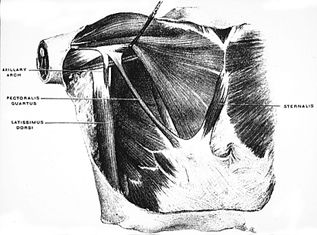
Axillary Arch .
from Huntington, 1905. — Anatomy Atlases. Immagine ospitata dal sito d’origine. Apri la pagina originale.
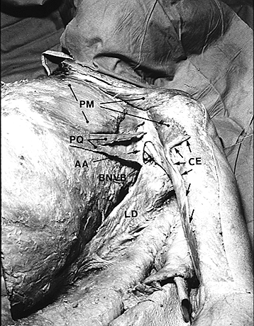
Axillary Arch, Pectoralis Quartus, Chondroepitrochlearis and the absence the twist of the tendon of Pectoralis Major.
from Bergman. — Anatomy Atlases. Immagine ospitata dal sito d’origine. Apri la pagina originale.
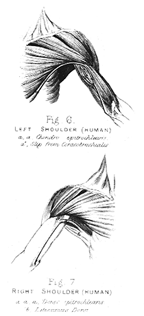
Chondroepitrochlearis (above) and Dorsoepitrochlearis (below)
from Perrin. — Anatomy Atlases. Immagine ospitata dal sito d’origine. Apri la pagina originale.
Riferimenti dell’opera originale
Elenco riportato come compare nella fonte, senza normalizzazione dei formati bibliografici.
- Alstead,S. (1933) Congenital defect of the pectoral muscles. Lancet (Lond.) 1:1179
- Bergman, R.A., Thompson, S.A., and F.A. Saadeh (1988) Anomalous fascicle and high origin of latissimus dorsi compensating for absence of serratus anterior. Anat. Anz. 167:161-164.
- Bergman, R.A. (1991) Doubled pectoralis quartus, axillary arch, chondroepitrochlearis, and the twist of the tendon of pectoralis major. Anat. Anz. 173:23-26.
- Birmingham, A. (1889) Homology and innervation of the achselbogen and pectoralis quartus, and the nature of the lateral cutaneous nerve of the thorax. J. Anat. Physiol. 23:206-223.
- Bryce, T.H. (1899) Note on the varieties of the pectoral sheet of muscle. J. Anat. Physiol. 34:75-78.
- Calori, L. (1866) Intorno ad alcune varietà incontrate nella muscolaturea degli arti superiori annotazioni anatomiche. Mem. de Accad. d. Scienze d. Istituto di Bologna. S. 2.VI:157-174.
- David, T.J. and R.M. Winter. (1985) Familial absence of pectoralis major, serratus anterior, and latissimus dorsi muscles. J. Med. Gen. 22:390-392.
- Dbaly, J. (1975) Ein Fall von beiderseitigem Vorkommen des muskulören Achselbogens. Anat. Anz. 137:75-78.
- Deville, -. (1848) Anomalie musculaire (grand pectoral et grand dorsal). Bulletins et Mem. de la Société Anatomique de Paris XXIII(1):319-320.
- Endres, H. (1893) Über ein Zwischenmuskelbündel im Gebiete des M. pectoralis major und latissimus dorsi. Anat. Anz. 8:387-397.
- Fitzgerald, R.R. (1935-36) A case showing the chondro-epitrochlearis muscle . J. Anat. 70:273-274.
- Gerard,G. et P. Cordier (1920) Au sujet d'un cas de fusion des tendons des muscles grand dorsal et grand round. Soc. Biol., Comptes Rendus Hebdomadaires des Séances et Mémoires. 83:927.
- Griffith, T.W. (1902) Note on a case of muscular abnormality observed during life. J. Anat. Physiol. 36:387-388.
- Henle, J. (1871) Handbuch der Muskellehre des Menschen, in Handbuch der systematischen Anatomie des Menschen. Verlag von Friedrich Vieweg und Sohn, Braunschweig.
- Kameda, Y. (1976) An anomalous muscle (accessory subscapularis-teres-latissimus muscle) in the axilla penetrating the brachial plexus. Acta Anat. 96:513-533.
- Kasai, T. and S.Chiba (1977) In Japanese. True nature of the muscular arch of the axilla and its nerve supply. Kaibogaku Zasshi 25:657-669.
- Langer, C. (1846) Zur Anatomie des Musculus latissimus dorsi. Oesterreich. Med. Wochenschrift. 1846:454-458, 486-492.
- LeDouble, A. (1891) Du muscle epitrochléo- olécranien et de sa signification au point de vue de l'anthropol. Paris. Bull. Soc. d'Anthrop. Paris.2:154-173.
- LeDouble, A. Les anomalies de muscle grand dorsal. Bull. Soc. d'Anthropol. Paris. 4:626-653.
- Macalister, A. (1875) Observations on muscular anomalies in the human anatomy. Third series with a catalogue of the principal muscular variations hitherto published. Trans. Roy. Irish Acad. Sci. 25:1-130.
- Miyauchi, R. (1982) A very rare variation of the latissimus dorsi muscle - A case with accessory insertion of the latissimus dorsi into the first rib and into the pectoralis major muscle. Okajimas Folia Anat. Jpn. 58:521-534.
- Mori, M. (1964) Statistics on the musculature of the japanese. Okajimas Folia Anatomica Japonica. 40:195-300.
- Mori, -. and -. Minoru. (1950) On the latissimus dorsi. Sotai Taiji no Kaibogakuteki Kenyu 11:1-37. In Japanese.
- Pichler, K. (1916) Ueber den Langer'schen Achselbogen. Anat. Anz. 49:310-318.
- Ramsay, A. (1812) An account of unusual conformation of some muscles and vessels. Edinburgh Med. Surg. J. 8:281-283.
- Regnault, F. (1902) Les causes des anomalies musculaires. L'Assoc. Anatomistes, Comptes Rendus. 4:19-20.
- Rao, G.R.K.H. and V.R. Rao. (1956) Dorso-epitrochlearis brachii. J. Anat Soc. India. 5:33-34.
- Wood, J. (1868) Variations in human myology observed during the winter session of 1867-68 at King's College, London. Proc. Roy. Soc. Lond. B 17:483-525.
- Note: See heading Axillary Arch for additional references. Panniculus carnosus and Pectoralis major for additional references.
Classificazione anatomica
- Sistemi:
- Sistema muscolare
Provenienza del contenuto
Adattamento
Adattamento di «Latissimus Dorsi — Illustrated Encyclopedia of Human Anatomic Variation: Opus I: Muscular System» di Ronald A. Bergman, Adel K. Afifi, Ryosuke Miyauchi. Fonte: https://www.anatomyatlases.org/AnatomicVariants/MuscularSystem/Text/L/06Latissimus.shtml Opera originale distribuita con licenza CC BY-NC-SA 4.0 (https://creativecommons.org/licenses/by-nc-sa/4.0/deed.it). Adattamento a cura di Redazione AIVA. Modifiche rispetto all’originale: Testo originale inglese riprodotto integralmente e riorganizzato nel formato scheda del portale (sintesi, testo, riferimenti), senza modificarne le affermazioni. La traduzione italiana non è ancora stata eseguita, e la pagina lo dichiara. Le figure non sono state copiate: sono collegate alla pagina d’origine. Questa pubblicazione non è approvata né avallata dagli autori dell’opera originale.
Licenza
Questo contenuto è distribuito con licenza CC BY-NC-SA 4.0. Licenze e attribuzioni