Voce enciclopedica
Levator Scapulae
Abstract
This muscle is composed of two parts that may vary independently of each other. Krause and Wood have classified these two as follows: A ventral fasciculus. This slip arises from the cervical vertebrae and runs downward and inserts on the ventral surface of subscapularis or serratus posterior superior. Other variations have been described : Macalister has found the following variations for the levator scapulae: Syn.: m. levator anguli scapulae, patientiae, Schulterheber, angulaire.
This muscle is composed of two parts that may vary independently of each other. Krause and Wood have classified these two as follows:
- A dorsal fasciculus, with two subtypes.
- the deviant fasciculus arises from the dorsal portion of the upper part of the origin (cervicle vertebrae) and runs downward and is inserted into serratus posterior superior, or the rhomboids, or onto a spinous process/es of vertebrae.
- a deviant fasciculus arises from the mastoid process, runs with splenius capitis and inserts onto the medial angle of the scapula.
A ventral fasciculus. This slip arises from the cervical vertebrae and runs downward and inserts on the ventral surface of subscapularis or serratus posterior superior.
Other variations have been described :
- a variable origin from the first to seventh cervical vertebrae, most commonly, however, it arises from the first and second. Mori reported that the origin, found most frequently in his series, was from cervicle 1, 2, 3, and 4 (66.6%). Cervicle 1, 2, and 3 was the next most common (26.6%). Cervicle 1 and 2 and Cervicle 1, 2, 3, 4, and 5 were less common origins (3.3%) for levator scapulae.
- Mori reported addlitional variations from his series. A deviant slip arises from the medial margin of levator scapulae, which runs medialward and downward to be inserted onto the spinous process of the second thoracic vertebra, or on the dorsal surface of serratus posterior superior, or on the dorsolumbar fascia. This variation was found by Mori in 9 (9%, right side 4 times and on the left 5 times) sides of 50 cadavers. In addition, Mori reported a deviant slip arising from the medial margin of levator scapulae, runs downward, and inserts on the ventral surface of subscapularis. This variation was found in 22 of 100 sides (on the right side in 7 cases and on the left in 15).
- slips may extend to the temporal or occipital bones.
- slips may extend to the rhomboids, serratus anterior, serratus posterior superior, and the trapezius muscles
- slips may extend to the clavicle, first and second ribs, and spinous process of the first thoracic vertebra
- parts of the muscle separated from each other, arising from a vertebral origin may insert on the angle of the scapula (levator anguli scapulae)
- a detached slip from various cervical vertebrae to the lateral end of the clavicle and acromion, corresponding to the levator claviculae found normally in other vertebrates; and
- insertion with rhomboideus minor, or bundles to splenius capitis or the scalenes may be found.
Macalister has found the following variations for the levator scapulae:
- The muscle may have a slip of origin from the squama temporis (Meckel, 1826)
- Or from the mastoid process (Blandin)
- Or from the trapezius
- With five separate vertebral origins, and one from the oblique process of the sixth cervical vertebra
- With a head from the second to the fourth dorsal spines
- Cleft into two parts, the upper coming from the first and second, the lower from the third and fourth cervical transverse processes (Wood); This splitting occurs in one of nine subjects, and is illustrated by Cuvier and Laurillard.
- Kelch (1813) found it cleft into three, and of these the middle was inserted into the scapulo-thoracic fascia
- Rosenmüller(1814) found a aberrant slip of this muscle passing from the first cervical transverse process into the first "tooth" of serratus magnus (anterior)
- A union of this muscle and serratus like that in the quadrumana (four- footed animals) is described by Henle
- A slip of levator was found to be inserted into the middle of serratus was found by Flower and Murie in a Bushwoman
- Wood found it split into six slips
- Macalister found it split into three parts
- In one case of fission into six, Wood found it to take origin from the fascia and fibers of the transversalis colli (the extension of longissimus into the neck, longissimus cervicis), the three upper and part of the fourth formed a normal muscle, the rest of the fourth and nearly all the fifth were inserted into the vertebral border of the scapula as far as the spine, intimately connected to the serratus magnus (anterior), the lowest slip was blended with rhomboideus minor at its insertion
- A slip onto the scapular spine was seen by M'Whinnie and Blandin
- Macalister found a separate slip lying deeper at first than the rest of the levator, finally crossing its insertion, to be inserted into the base of the triangular space at the root of the spine of the scapula
- A slip missing the scapula, and inserted between it and the spinal column, was seen by Otto and Meckel; a similar slip inserted into the second rib was seen by Blandin, Meckel, and Theile; in another case, Clason found a lower part of levator. inserted onto the first and second ribs anterior to serratus magnus (anterior)
- It has been found sending a slip to be inserted with the serratus posticus superior (Theile and Kelch)
- Or to the scalenus posticus
- Or scalenus medius (Wood)
- to splenius capitis (Wood)
- Or to complexus (semispinalis capitis).
Syn.: m. levator anguli scapulae, patientiae, Schulterheber, angulaire.

Varieties of Shoulder Muscles-Levator Scapulae.
- Modified and redrawn from Wood.
Anatomy Atlases. Immagine ospitata dal sito d’origine. Apri la pagina originale.
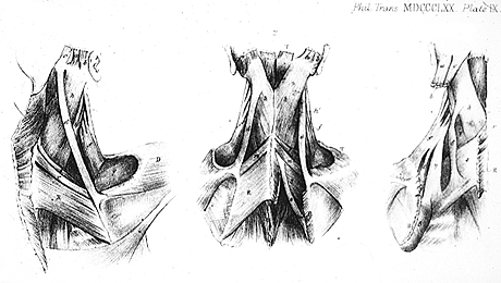
Muscles of the Scapula, Neck, and Head.
- Left 1, Back view of the human shoulder, showing occipito-scapularis (d) on the right side.
- Middle 2, Back view on both sides, showing varieties homologous with the occipito- scapularis (d,d), and one other anomalous slip (ø) of rhomboideus major (R).
- Right 3, Back view on left side, showing homologous occipito-scapularis (�) and two slips (e and n) from levator scapulae (f) to serratus anterior (S) and serratus posterior superior (s).
from Wood. — Anatomy Atlases. Immagine ospitata dal sito d’origine. Apri la pagina originale.
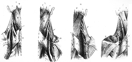
Muscles of the Scapula, Neck, and Head.
- Left 4, Back view on left side, showing slips (e and n) of levator scapulae (f) to serratus posterior superior (s) and serratus anterior (S).
- Left middle 5, Back view on right side, showing homologous slips (e and n) to the same muscles from the front surface of levator scapulae (f).
- Right middle 6, Back view on right side, showing homologous slip (n) in front of levator scapulae (f) to serratus anterior (S).
- Right 7. Back view on left side, showing slips (F) of levator scapulae (f) to serratus anterior (S) and rhomboideus minor (r).
from Wood. — Anatomy Atlases. Immagine ospitata dal sito d’origine. Apri la pagina originale.
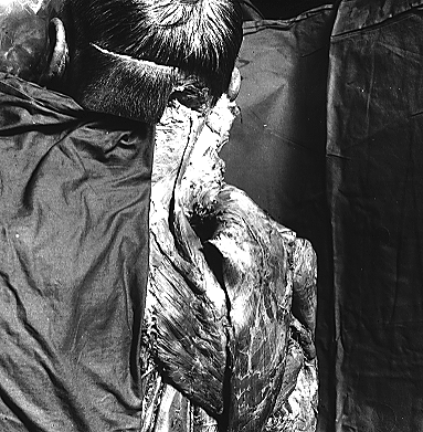
Occipitoscapularis.
- Dissecting room specimen,
University of Iowa. — Anatomy Atlases. Immagine ospitata dal sito d’origine. Apri la pagina originale.
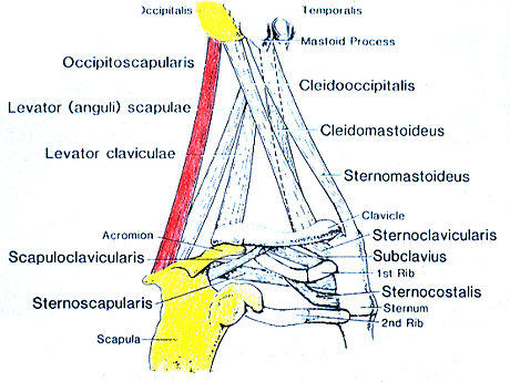
Occipitoscapularis.
Redrawn and modified from Wood. — Anatomy Atlases. Immagine ospitata dal sito d’origine. Apri la pagina originale.
Riferimenti dell’opera originale
Elenco riportato come compare nella fonte, senza normalizzazione dei formati bibliografici.
- Anson, B.J., Ed. (1966) Morris' Human Anatomy,12th ed., The Blakiston Division, McGraw-Hill Book Company, New York
- Henle, J. (1871) Handbuch der Muskellehre des Menschen, in Handbuch der systematischen Anatomie des Menschen. Verlag von Friedrich Vieweg und Sohn, Braunschweig.
- Latarjet, A. (1948) Testut's Traité D'Anatomie Humaine, 9th ed., G. Doin & Cie., Paris.
- Macalister, A. (1875) Additional observations on muscular anomalies in human anatomy (third series), with a catalogue of the principal muscular variations hitherto published. Trans. Roy. Irish Acad. Sci. 25:1-134.
- Mori, M. (1964) Statistics on the musculature of the Japanese. Okajimas Fol. Anat. Jap. 40:195-300.
- Mori, -. and -. Minoru. (1950) On the M. levator scapulae. Sotai Taiji no Kaibogakuteki Kenkyu. 11:79-153. In Japanese.
- Schaefer, E.A., Symington, J. and T.H. Bryce, Eds. (1923) Quain's Anatomy. 11th ed. Longmans, Green and Co., London.
- Tochiuchi, I. and K. Tsukahara. (1944) On the anomaly of the M. levator scapulae. Iwate Ikakusenmongakko Zasshi. 7:36-92. In Japanese.
- Wood, J. (1867) On human muscular variations in their relation to comparative anatomy. J. Anat. Physiol. 15: 44-59.
- Wood, J. (1868) Variations in human myology observed during the winter session of 1867-68 at king's College, London. Proc. Roy. Soc. Lond. B. 17: 483-525.
- Wood, J. (1870) On a group of varieties of the muscles of the human neck, shoulder, and chest, and their transitional forms and homologies in the mammalia. Phil. Trans. Roy. Soc. Lond. 160:83-116.
- Yamada, K. (1933) One case of the variation of the M. levator scapulae. Aichi Igakkai Zasshi. 40:1351-1354.
- Yamada, K. (1934) Ein fall von Variation des M. levator scapulae beim Menschen. Nagoya Journal of Medical sciences.
Classificazione anatomica
- Sistemi:
- Sistema muscolare
Provenienza del contenuto
Adattamento
Adattamento di «Levator Scapulae — Illustrated Encyclopedia of Human Anatomic Variation: Opus I: Muscular System» di Ronald A. Bergman, Adel K. Afifi, Ryosuke Miyauchi. Fonte: https://www.anatomyatlases.org/AnatomicVariants/MuscularSystem/Text/L/14Levator.shtml Opera originale distribuita con licenza CC BY-NC-SA 4.0 (https://creativecommons.org/licenses/by-nc-sa/4.0/deed.it). Adattamento a cura di Redazione AIVA. Modifiche rispetto all’originale: Testo originale inglese riprodotto integralmente e riorganizzato nel formato scheda del portale (sintesi, testo, riferimenti), senza modificarne le affermazioni. La traduzione italiana non è ancora stata eseguita, e la pagina lo dichiara. Le figure non sono state copiate: sono collegate alla pagina d’origine. Questa pubblicazione non è approvata né avallata dagli autori dell’opera originale.
Licenza
Questo contenuto è distribuito con licenza CC BY-NC-SA 4.0. Licenze e attribuzioni