Voce enciclopedica
Maxilla
Abstract
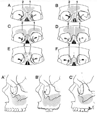
In the maxilla, the inferior wall of the infraorbital canal may be incomplete, and hence the infraorbital nerve is in contact with the mucosa of the maxillary sinus. In about 5% of skulls, the infraorbital groove is present as a canal. Two or more infraorbital foramina have been reported. The infraorbital foramen may lie anywhere from 3 to 7 mm below the infraorbital margin. Occasionally, a bony spine covers the foramen. The infraorbital canal (groove) may be shifted laterally as far as the suture between the maxilla and zygomatic bones.
In the maxilla, the inferior wall of the infraorbital canal may be incomplete, and hence the infraorbital nerve is in contact with the mucosa of the maxillary sinus. In about 5% of skulls, the infraorbital groove is present as a canal. Two or more infraorbital foramina have been reported. The infraorbital foramen may lie anywhere from 3 to 7 mm below the infraorbital margin. Occasionally, a bony spine covers the foramen. The infraorbital canal (groove) may be shifted laterally as far as the suture between the maxilla and zygomatic bones. Supplementary infraoribital canals sometime occurs medial, lateral and/or superior to the usual canal.
The earliest account of variation in the infraorbital canal was given by Gruber (1875). Gruber reported that the number of foramina may vary from 1 to 5. It was Kadanoff, Mutafov and Jordanov in 1970, that tabulated and illustrated the variety of infraorbital foramina that were found in over fourteen hundred skulls. Kadanoff, et al. found it doubled in 131 (9%), tripled in 7 (0.5%), and greater than three in 4 (0.3).
Berry in 1975 reported accessory infraorbital foramina in skulls from four geographic areas: English, 2.2% in men and 4.8% in women" Burmese, 6.4% in men and 8.7% in women; Americans (Northwest), 12.5% in men and 7.9% in women; Mexican, 18.2% in men and 12.5% in women. The numbers of accessory foramina were not discussed. Two hundred-fifty skulls (500 half skulls) in the Iowa collection did not have more than one foramen per half skull but two were divided by a partition.
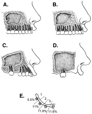
Investigations have shown that enlargement of the maxillary sinus may be produced by (a) hollowing out of the alveolar process (alveolar recess); (b) excavation of the floor of the nasal fossa by extension of the alveolar recess between the plates of the hard palate (palatal recess); (c) encroachment of the sinus into the frontal process of the maxilla; (d) hollowing out of the zygomatic process of the zygomatic bone (malar recess); and (e) extension to and appropriation of an air cell within the orbital process of the palatine bone (palatal recess). Reduction of the maxillary sinus, on the other hand, may follow (a) imperfect absorption of cancellous bone on the floor of the sinus, or secondary thickening of its walls; or (b) encroachment due to approximation of the facial and nasal walls, unusual depression of the canine fossa, excessive bulging of the lateral nasal wall, or imperfectly erupted teeth. The crescentric projections that quite commonly protrude from the walls into the interior occasionally are replaced by septa that completely divide the sinus into two cavities, each having an independent opening into the nasal fossa, but having no communication with each other. These partitions vary in position and direction, in some cases subdividing the sinus into an anterior and a posterior compartment, and in others, into an upper and a lower chamber. In the latter case, the lower chamber may communicate with the inferior meatus.
Accessory ostia of the maxillary sinus are found in about 30% of skulls; as many as three in one skull have been reported.
The incisive part of the alveolar process may be an independent bone, the os incisivum. The incisive canals, which permit passage of the nasopalatine nerves, may open by several foramina.
Deficiency of the palatine processes result in cleft palate. Separate bone may occur on either side of the median palatine suture. The palatine processes of the maxilla sometimes project backward in the median plane, separating the horizontal plates from each other.
In the suture between the ethmoid and maxilla, beneath the anterior ethmoidal foramen, a 3 mm hiatus may occur through which the orbital and nasal cavities communicate.
The maxillary sinus was first described and illustrated by Leonardo da Vinci in 1489.
According to Kadanoff (1970), 90% of infraorbital foramina are single; 9% are doubled, and 0.05% are tripled in number.
Variations in Size of Frontal and Maxilary Sinuses .
Anatomy Atlases. Immagine ospitata dal sito d’origine. Apri la pagina originale.
Anatomy Atlases. Immagine ospitata dal sito d’origine. Apri la pagina originale.
Riferimenti dell’opera originale
Elenco riportato come compare nella fonte, senza normalizzazione dei formati bibliografici.
- Berry, A.C. (1975) Factors affecting the incidence of non-metrical skeletal varients. J. Anat. 120:519-535.
- Boberg, J. Barber, A. and S.S. Barber. (1980) Neural blockade for Ophthalmologic Surgery. In Cousins, M.J. and P.O. Brodenbaugh, eds. Neural Blockage in Clinical Anaesthesia and Management of Pain. J.B. Lippincott Co., Philadelphia.
- Canneyt, M.J.G. (1945) Dent dans la cavité orbitaire. Bull. Soc. Belge Ophthalmol. 82:123-127. Cited in Excerpta Medica, Sec. 1, Vol. 2. abstract 191, p. 97, 1948.
- Derry, D.E. (1937-38) Two skulls with absence of the premaxilla. J. Anat. 72:295-298.
- D'Este, L.S. (1900) Les incisures, les trous et les canaux sus-orbitares avec leur nerfs respectifs, et la résecttion du nerf sus-orbitaire. Arch. Ital. Biol. 33:481.
- Dubreuil-Chambardel, L. (1920) Los fonto-lacrymo-naso-maxillare. Bull. et Mém. de la Soc. d'Anthrop. de Paris 1:35-36.
- DuBrul, L.E. (1988) Sicher and DuBrul's Oral Anatomy, 8th ed., Ishiyaku EuroAmerica, Inc., St. Louis.
- Gozdziewski, S., Nizankowski, C. and R. Kindlik. (1979) Die morphologische Analyse des Canalis infraorbitalis und des Foramen infraorbitale beim Menschen. Anat. Anz. 145:517-527.
- Gruber, W. (1873) Über eingige merkwürdige Oberkiefer Abweichungen. Arch. Anat. Physiol. Wissen. Med. 1873:195-207.
- Gruber, W. (1874)Über die infraorbitalen Kanäle des Menschenund Säugetiere. Mem. Acad. Imp. Sci. St. Petersburg 21:1-27
- Gruber, W. (1878) Ein hakenförmiges Fortsätzchen über und vor dem Infraorbitalloche. Arch. Pathol. Anat. Physiol. Klin. Med. 72:494-496.
- Gruber, W. (1888) Über Fälle von Teilung des Sinus maxillaris durch ein Septum osseum perfuctum in zwei von einander völlig abgeschlossene Sinus maxillaris secundarii mit separaten öffnungen in den Meatus narium medius. Arch. Pathol. Anat. Physiol. Klin. Med. 113:530-533.
- Gruber, W. (1874) Über die infraorbitalen Kanale des Menschen und der Saugetierre. Mem. Akad. Imp. Sci. St. Petersburg. 21(10):1-27.
- Herpin, A. (1926) Des anomalies maxillo-dentaires. Assoc. Anatomistes Comptes Rendus 21:270-272.
- Jordan, J. (1967) Double infraorbital nerve in man. Folia Morphologica 26(4):405-407.
- Kadanoff, D., Mutafov, St. and J. Jordanov. (1970) Über die Hauptöffnungen resp. Incisurae des Gesichtsschädels. Morphologisches Jahrbuch 115:102-118.
- Koerbin, -. (1878) Anomale Nahtbildung am Oberkiefer-Stirnforsatz. Arch. Pathol. Anat. Physiol. Klin. Med. 73:121-125.
- Kurlej, W., Gozdziewski, S. and J. Marek. (1983) Morphology of the incisive fossa, canal and foramen in man. Folia Morphol. (Warsaw) 42:129-138.
- Lang, J. (1983) Clinical Anatomy of the Head. Neurocranium; Orbit; Craniocervical Regions. Springer-Verlag, Berlin, Heidelberg, New York.
- Leo, J., Cassell, M.D. and R.A. Bergman (1995) Variation of human infraorbital nerve, canal, and foramen. Ann. Anat. 177:(1):93-95.
- Myerson, M.C. (1932) The natural orifice of the maxillary sinus. 1. Anatomic studies. Arch. Otolaryngol. 15:80-91.
- Rosenberger, H.C. (1938) The clinical availability of the ostium maxillare: A clinical and cadaver study. Ann. Otol., Rhin. Laryng. 47:176-182.
- Roujou, A. (1876) Sur la persistence des intermaxillaires chez l'homme. Bull. de la Soc. d'Anthrop. de Paris. 11:171-172.
- Schultz, A.H. (1954) Die Foramina infraorbitalia bei Primates. Z. Morph. Anthrop. 46:404- 407.
- Sedwick, H.J. (1934) Form, size and position of the maxillary sinus at various ages studied by means of roentgenograms of the skull. Am. J. Roentgenol. 32:154-160.
- Simon, E. (1939) Anatomy of the opening of the maxillary sinus. Arch. Otolaryngol. 29:640-649.
- Thomson, A. (1890) The orbito-maxillary frontal suture in man and apes, with notes on the varieties of the human lachrymal bone. J. Anat. Physiol. 24:349-357.
- Tondury, G. (1946) Zur Genese der Hasenscharte beim Menschen. Rev. Suisse Zool. 53:459-468. Cited in Excerpta Medica, Sec 1, Vol 2, abstract 299, p. 146, 1948.
- Underwood, A.S. (1910) An inquiry into the anatomy and pathology of the maxillary sinus. J. Anat. Physiol. 44:354-369.
- Van Alyea, O.E. (1936) The ostium maxillare: Anatomic study of its surgical accessibility. Arch. Otolaryng. 24:553-569.
- von Wendler, D. (1986) Nathanael Highmore (1613-1685) und die Oberkiefhöhle. Anat. Anz. 162:272-280.
- Vram, U. (1899) Sur un os interstitiel naso-maxillaire dans un crâne humain. Arch. Ital. Biol. 32:455.
- Walker, C. (1917) Absence of pre-maxilla. J. Anat. 51:392-395.
- Wendler, Von D. (1986) Nathanael Highmore (1613-1685) und die Oberkieferhöhle Anat. Anz. 162:375-380.
- Williams, P.L., Warwick, R., Dyson, M. and L.H. Bannister. (1989) Gray's Anatomy, 3 /1h ed., Churchill and Livingstone, Edinburgh.
Classificazione anatomica
- Sistemi:
- Sistema scheletrico
Provenienza del contenuto
Adattamento
Adattamento di «Maxilla — Illustrated Encyclopedia of Human Anatomic Variation: Opus I: Muscular System» di Ronald A. Bergman, Adel K. Afifi, Ryosuke Miyauchi. Fonte: https://www.anatomyatlases.org/AnatomicVariants/SkeletalSystem/Text/Maxilla.shtml Opera originale distribuita con licenza CC BY-NC-SA 4.0 (https://creativecommons.org/licenses/by-nc-sa/4.0/deed.it). Adattamento a cura di Redazione AIVA. Modifiche rispetto all’originale: Testo originale inglese riprodotto integralmente e riorganizzato nel formato scheda del portale (sintesi, testo, riferimenti), senza modificarne le affermazioni. La traduzione italiana non è ancora stata eseguita, e la pagina lo dichiara. Le figure non sono state copiate: sono collegate alla pagina d’origine. Questa pubblicazione non è approvata né avallata dagli autori dell’opera originale.
Licenza
Questo contenuto è distribuito con licenza CC BY-NC-SA 4.0. Licenze e attribuzioni
