Voce enciclopedica
Spinal Cord
Abstract
Diastematomyelia is a condition in which a portion of or the entire spinal cord is divided into two lateral halves. Each half of the cord is enveloped in its own membranes and give rise to its own spinal roots. Hydromyelia or hydrorrhachis interna is a defective closure or arrangement of the divisions of the primary fetal central canal. Its presence may be indicated by subsequent development of syringomyelia or by its association with spina bifida. Hydrorrhachis externa is the abnormal congenital accumulation of fluid between the meninges of the cord causing a diminution of cord volume.
Diastematomyelia is a condition in which a portion of or the entire spinal cord is divided into two lateral halves. Each half of the cord is enveloped in its own membranes and give rise to its own spinal roots.
Hydromyelia or hydrorrhachis interna is a defective closure or arrangement of the divisions of the primary fetal central canal. Its presence may be indicated by subsequent development of syringomyelia or by its association with spina bifida. Hydrorrhachis externa is the abnormal congenital accumulation of fluid between the meninges of the cord causing a diminution of cord volume.
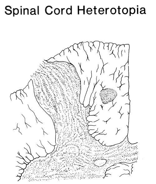
Heterotopias (misplacement of parts) of the spinal cord include gray matter islands located in the white matter; these may be isolated or partially joined to the gray horns by slender strands of gray matter.
Malposition of bundles of white matter or passage of bundles of white matter (nerve fibers) through unusual sites to reach their destination has been described. Additional variations include: (a) doubling of the spinal cord in some of its parts; (b) the spinal cord (in an adult) extending to the level of the third sacral vertebra; and (c) doubling of the central canal. The denticulate ligaments vary in number between 18 and 22 in normal individuals.
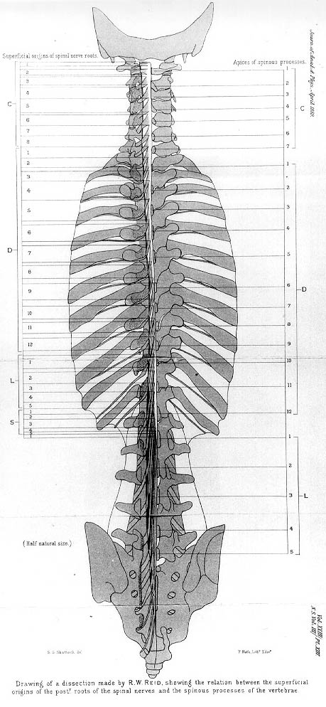
In one study of 129 adults, the point of termination of the spinal cord varied from the lower third of the twelfth thoracic vertebra to the middle third of the third lumbar. In 95% of these 129 cases, the cord terminated at the level of the first and second lumbar vertebra.
Anatomy Atlases. Immagine ospitata dal sito d’origine. Apri la pagina originale.

Anatomy Atlases. Immagine ospitata dal sito d’origine. Apri la pagina originale.
Anatomy Atlases. Immagine ospitata dal sito d’origine. Apri la pagina originale.
Riferimenti dell’opera originale
Elenco riportato come compare nella fonte, senza normalizzazione dei formati bibliografici.
- Holiday, M. and R.P. Cormack. (1927) Notes on a case of partial duplication of the spinal cord in the lumbar region. Trans R. Soc. Trop. Med. Hygiene 21:215-218.
- Jit, I. and V.M. Charnalia. (1959) The vertebral level of the termination of the spinal cord. J. Anat. Soc. India 8:93-101.
- Joylet, -. (1867) Sur un cas d'anomalie du canal central de la moelle épinière, observée sur la moelle de Mme. V., agée de 80 ans, morte à la Salpêtière. Soc. Biol. Comptes Rendus des Séances et Mémoires 19:7-8.
- Kraus, W. and A. Weil. (1926) Duplicité de la moelle Epinière. Rev. Neurol. 33:317-323.
- Lazorthes, G., Poulhès, J. et S. Gaubert. (1952) La significance et la hauteur des arcades du ligament dentelé. Assoc. Anatomistes Comptes Rendus 39:303-311.
- Reimann, A.F. and B.J. Anson. (1944) Vertebral level of termination of the spinal cord with report of a case of sacral cord. Anat. Rec. 88:127-138.
- Trétiakoff, C. et V. Ramos. (1921) Etude anatomique d'un cas de moelle bifide (double moelle lombaire). Rev. Neurol. 28:768-772.
- Van Gieson, I. (1892) A study of the artifacts of the nervous system. N.Y. Med. J. 56:337-447.
- Wagner, J. (1861) Notiz über einen teilweise doppelten Centralkanal im Rückenmark des Menschen. Arch. Anat. Physiol. Wissen. Med. 1861:735-747.
- Walker, A.E. (1944) Dilatation of the vertebral canal associated with congenital anomalies of the spinal cord. Am. J. Roentgenology 52:571-582.
Classificazione anatomica
- Sistemi:
- Sistema nervoso
Provenienza del contenuto
Adattamento
Adattamento di «Spinal Cord — Illustrated Encyclopedia of Human Anatomic Variation: Opus I: Muscular System» di Ronald A. Bergman, Adel K. Afifi, Ryosuke Miyauchi. Fonte: https://www.anatomyatlases.org/AnatomicVariants/NervousSystem/Text/SpinalCord.shtml Opera originale distribuita con licenza CC BY-NC-SA 4.0 (https://creativecommons.org/licenses/by-nc-sa/4.0/deed.it). Adattamento a cura di Redazione AIVA. Modifiche rispetto all’originale: Testo originale inglese riprodotto integralmente e riorganizzato nel formato scheda del portale (sintesi, testo, riferimenti), senza modificarne le affermazioni. La traduzione italiana non è ancora stata eseguita, e la pagina lo dichiara. Le figure non sono state copiate: sono collegate alla pagina d’origine. Questa pubblicazione non è approvata né avallata dagli autori dell’opera originale.
Licenza
Questo contenuto è distribuito con licenza CC BY-NC-SA 4.0. Licenze e attribuzioni