Voce enciclopedica
Supinator
Abstract
The division of the muscle into two lamellae may be more pronounced than is normally the case. An accessory fasciculus from the lateral epicondyle has been observed, as well as fibers inserted into the tendon of insertion of the biceps, the bursa under the tendon, and the tuberosity of the radius. Spinner (1968) has pointed out that the most proximal part of the superficial head of the supinator muscle may be tendinous and form a fibrous arch and is implicated in the paralysis of the posterior interosseous nerve.
The division of the muscle into two lamellae may be more pronounced than is normally the case. An accessory fasciculus from the lateral epicondyle has been observed, as well as fibers inserted into the tendon of insertion of the biceps, the bursa under the tendon, and the tuberosity of the radius.
Spinner (1968) has pointed out that the most proximal part of the superficial head of the supinator muscle may be tendinous and form a fibrous arch and is implicated in the paralysis of the posterior interosseous nerve. The posterior interosseous nerve passes in a plane between the two heads of the supinator beneath the arch forming the origin of the muscle. The arch arises in a semicircular manner from the tip of the lateral epicondyle, its fibers dip downwards about 1cm. and attaches to the medial aspect of the lateral epicondyle just lateral to the articular surface of the capitulum. The posterior interosseous nerve passes beneath the edge of the fibrous arch where it may become entrapped or compressed. In adults specimens, there is considerable variation in the thickness of the fibrous arch and in the size of the opening for the interosseous nerve. In addition to its origin from the radial notch (lesser sigmoid) notch of the ulna, the superficial head of the supinator arises from the lateral aspect of the lateral epicondyle. In 70% of specimens dissected, Spinner found the medial half of the arcade was membranous and pliable, being inserted into the medial aspect of the lateral epicondyle adjacent to the capitulum. In the remaining 30% the medial half of the arcade was the same firm, unyielding, tendinous consistency as the lateral half. This fibrous arcade of Frohse was present in all ten cases of posterior interosseous nerve paralysis treated by Spinner.
Macalister reports the following variations in supinator: 1. The muscle may be doubled (Sandifort); 2. A cleft may be found where the posterior interosseus nerve perforates it; 3. A slip may insert on the tubercle of the radius; 4. A similar slip was found joining pronator teres (Clason); 5. A second or supinator brevis accessorius (Halbertsma) was found. It is a slip from the lower border of brachialis inserted onto the tubercle of the radius; 6. Dursey described a slip from this muscle inserted into the biceps tendon, and a slip into the bursa tubercularis of the radius.
See also brachioradialis .
Syn.: m. supinator brevis (Henle), Kurzer Rückwärtswender.
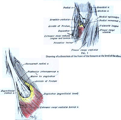
Arcade of Frohse.
from Spinner — Anatomy Atlases. Immagine ospitata dal sito d’origine. Apri la pagina originale.
Reference Anson, B.J., Ed. (1966) Morris' Human Anatomy,12ed., The Blakiston Division, McGraw-Hill Book Company, New York.
Carrington, R.E., Horrocks, P. and W.H. White. (1883) Abnormalities observed in the dissecting room of Guy's hospital during the sessions 1880-81 and 1881-82. Guy's Hosp. Reports. 46:57-81. (Volume 26 of the third series).
Clason, E. (1869) Om muskelanomalier. Upsala Lakareforening Forhandlingar. IV:244-248.
Frohse, F. and M. Fränkel. (1908) Die Muskeln des Menschlichen Armes. Jena.
Halbertsma, H.J. (1862) Ontleedkundige Aanteekeningen. Verslagen en Medadeelingen der Koninklijke Akademie van Wetenschappen, Afdeeling Natuurkunde. 13:330-347.
Macalister, A. (1875) Observations on the muscular anomalies in the human anatomy. Third series with a catalogue of the principal muscular variations hitherto published. Trans. Roy. Irish Acad. Sci. 25:1-130.
Spinner, M. (1968) The arcade of Frohse and its relationship to posterior interosseous nerve paralysis. J. Bone Joint Surg. B 50:809-881.
Classificazione anatomica
- Sistemi:
- Sistema muscolare
Provenienza del contenuto
Adattamento
Adattamento di «Supinator — Illustrated Encyclopedia of Human Anatomic Variation: Opus I: Muscular System» di Ronald A. Bergman, Adel K. Afifi, Ryosuke Miyauchi. Fonte: https://www.anatomyatlases.org/AnatomicVariants/MuscularSystem/Text/S/53Supinator.shtml Opera originale distribuita con licenza CC BY-NC-SA 4.0 (https://creativecommons.org/licenses/by-nc-sa/4.0/deed.it). Adattamento a cura di Redazione AIVA. Modifiche rispetto all’originale: Testo originale inglese riprodotto integralmente e riorganizzato nel formato scheda del portale (sintesi, testo, riferimenti), senza modificarne le affermazioni. La traduzione italiana non è ancora stata eseguita, e la pagina lo dichiara. Le figure non sono state copiate: sono collegate alla pagina d’origine. Questa pubblicazione non è approvata né avallata dagli autori dell’opera originale.
Licenza
Questo contenuto è distribuito con licenza CC BY-NC-SA 4.0. Licenze e attribuzioni