Voce enciclopedica
Supraclavicular Nerves
Abstract
The following variations have been reported in these nerves. A branch to the phrenic nerve may pass down into the thorax over the subclavian artery or vein before joining the phrenic nerve. An additional bundle from the fifth cervical nerve joining this branch has been reported. A branch to the rhomboid muscles may pass through the trapezius muscle on its way to the skin close to the midline, at the level of the spinous processes of the fifth and sixth thoracic vertebrae. In 10 of 254 cases, the clavicle was perforated by the middle branch of the supraclavicular nerves.
The following variations have been reported in these nerves. A branch to the phrenic nerve may pass down into the thorax over the subclavian artery or vein before joining the phrenic nerve. An additional bundle from the fifth cervical nerve joining this branch has been reported.
A branch to the rhomboid muscles may pass through the trapezius muscle on its way to the skin close to the midline, at the level of the spinous processes of the fifth and sixth thoracic vertebrae.
In 10 of 254 cases, the clavicle was perforated by the middle branch of the supraclavicular nerves. They traversed the bone in intraclavicular canals and were visible on x-ray films.
Image 31A Image 31B Image 31C Image 31D Image 31E Image 31F
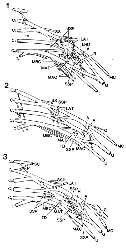
Anatomy Atlases. Immagine ospitata dal sito d’origine. Apri la pagina originale.
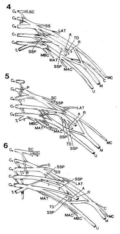
Anatomy Atlases. Immagine ospitata dal sito d’origine. Apri la pagina originale.
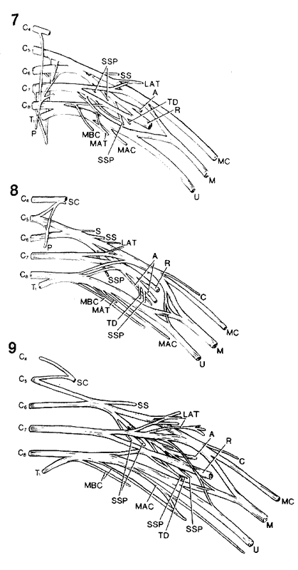
Anatomy Atlases. Immagine ospitata dal sito d’origine. Apri la pagina originale.
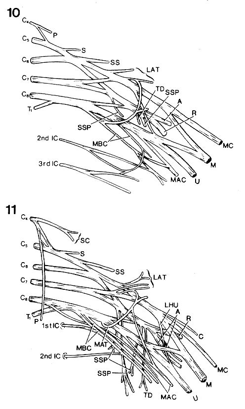
Anatomy Atlases. Immagine ospitata dal sito d’origine. Apri la pagina originale.
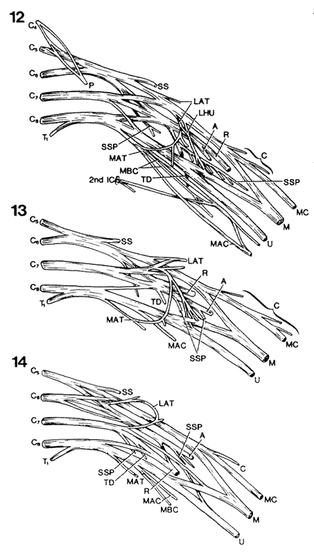
Anatomy Atlases. Immagine ospitata dal sito d’origine. Apri la pagina originale.
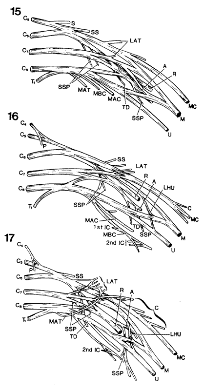
Anatomy Atlases. Immagine ospitata dal sito d’origine. Apri la pagina originale.
Image 31G Image 31H Image 31I Image 31J
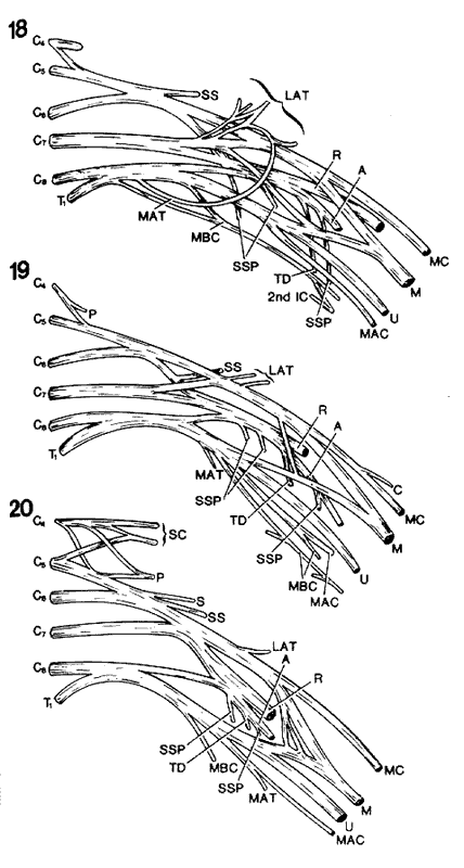
Anatomy Atlases. Immagine ospitata dal sito d’origine. Apri la pagina originale.
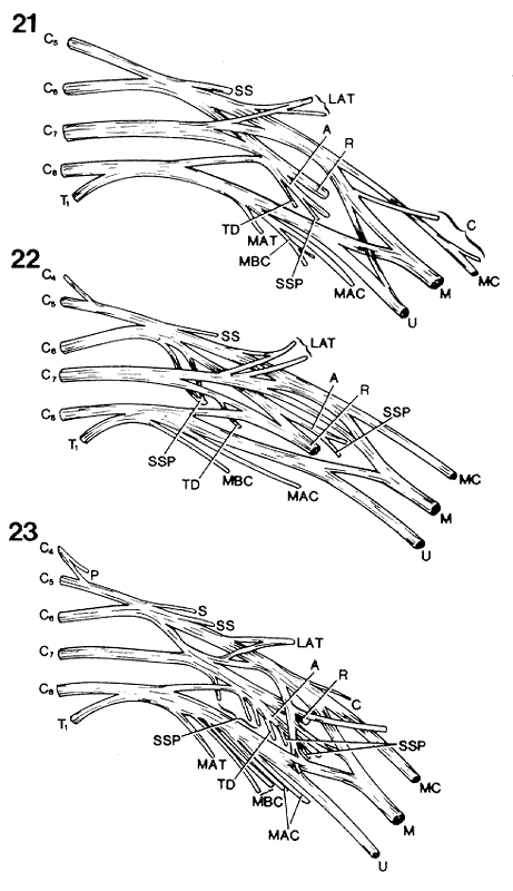
Anatomy Atlases. Immagine ospitata dal sito d’origine. Apri la pagina originale.
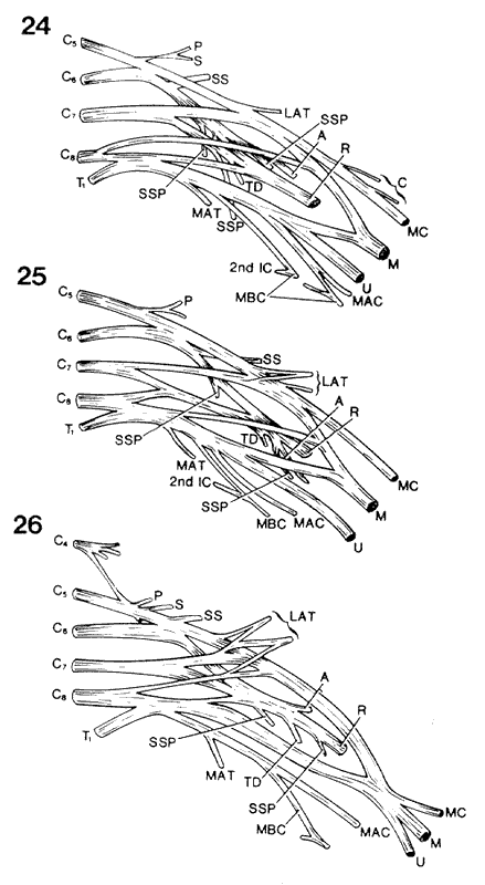
Anatomy Atlases. Immagine ospitata dal sito d’origine. Apri la pagina originale.
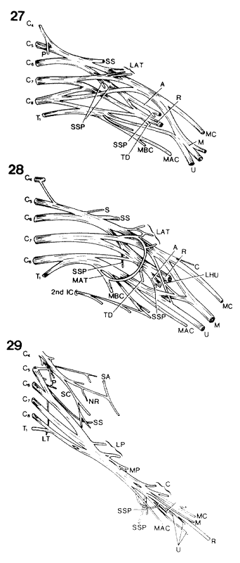
Anatomy Atlases. Immagine ospitata dal sito d’origine. Apri la pagina originale.
Riferimenti dell’opera originale
Elenco riportato come compare nella fonte, senza normalizzazione dei formati bibliografici.
- Anson, B.J., Ed. (1966) Morris' Human Anatomy, 12th ed. The Blakiston Division, McGraw-Hill Book Company, New York.
- Henle, J. (1868) Handbuch der Systematischen Anatomie des Menschen, von Friedrich Vieweg und Sohn, Braunschweig.
- Kopsch, F. (1908) Rauber's Lehrbuch der Anatomie des Menschen. Georg Thieme, Leipzig.
- Latarjet, A. (1948) Testut's Traité d'Anatomie Humaine, 9th ed., G. Doin & Cie., Paris.
- Olivier, E., Minne, -. et -. Rocquet. (1926) La traversée diaphragmatique du nerf phrenique droit et ses variations. Assoc. Anatomistes Comptes Rendus 21:441-444.
- Papadatos, D. (1980) Supraclavicular nerves piercing the clavicle: A study of 10 cases. Anat. Anz. 147:371-381.
Classificazione anatomica
- Sistemi:
- Sistema nervoso
Provenienza del contenuto
Adattamento
Adattamento di «Supraclavicular Nerves — Illustrated Encyclopedia of Human Anatomic Variation: Opus I: Muscular System» di Ronald A. Bergman, Adel K. Afifi, Ryosuke Miyauchi. Fonte: https://www.anatomyatlases.org/AnatomicVariants/NervousSystem/Text/SupraclavicularNerves.shtml Opera originale distribuita con licenza CC BY-NC-SA 4.0 (https://creativecommons.org/licenses/by-nc-sa/4.0/deed.it). Adattamento a cura di Redazione AIVA. Modifiche rispetto all’originale: Testo originale inglese riprodotto integralmente e riorganizzato nel formato scheda del portale (sintesi, testo, riferimenti), senza modificarne le affermazioni. La traduzione italiana non è ancora stata eseguita, e la pagina lo dichiara. Le figure non sono state copiate: sono collegate alla pagina d’origine. Questa pubblicazione non è approvata né avallata dagli autori dell’opera originale.
Licenza
Questo contenuto è distribuito con licenza CC BY-NC-SA 4.0. Licenze e attribuzioni