Voce enciclopedica
Osso temporale
Abstract
L’osso temporale varia per persistenza di suture fetali, estensione e asimmetria delle cellule mastoidee, forma del processo mastoideo e del canale carotico. Il processo stiloideo varia marcatamente in lunghezza, può essere composto da più ossicini o mancare, e anche gli ossicini dell’udito mostrano variazioni.
È stata riportata l’assenza della parte superiore della squama temporalis.
Si riscontra frequentemente una sutura frontosquamosa, che si trova normalmente nei crani dello scimpanzé, del gorilla e del gibbone.
La sutura petrosquamosa dell’osso temporale talvolta persiste. Le cellule mastoidee variano per estensione e possono invadere la parte squamosa dell’osso; le cellule pneumatiche possono non essere simmetriche sui due lati.
La squama in alcuni casi è divisa in due o quattro parti.
Una barra ossea nella dura al di sopra del nervo trigemino è stata interpretata come un vestigio della parete cranica primitiva quale è presente nei rettili.
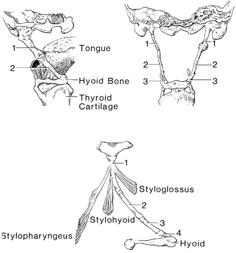
I processi stiloidei variano marcatamente in lunghezza e il legamento stiloioideo può calcificarsi realizzando una connessione rigida con l’osso ioide. È stato riportato un processo stiloideo lungo 75 mm. Il processo stiloideo può essere composto da due a cinque ossicini, articolati fra loro mediante sincondrosi.
Il grande corno dello ioide può essere unito al corpo dell’osso ioide mediante un’articolazione. L’arco zigomatico può essere sostituito da una banda fibrosa. In alcuni casi il processo zigomatico dell’osso temporale è assente.
Il processo mastoideo può possedere un doppio apice, con la porzione mediale separata dalla laterale da una fessura. Di solito il labbro mediale del solco forma una cresta distinta e offre il punto di attacco del muscolo digastrico; un processo paramastoideo ingrandito può risultare dallo sviluppo di cellule pneumatiche all’interno della porzione mediale del processo. Le cellule pneumatiche, di norma limitate al processo mastoideo, possono invadere la parte orizzontale o verticale della squama, o persino la pars petrosa.
Le cellule mastoidee variano grandemente per estensione e la pneumatizzazione può essere asimmetrica sui due lati.
La pars petrosa è molto raramente variabile. La fossa subarcuata, tuttavia, può persistere, in parte, come canale osseo. Nei casi di assenza dell’arteria carotide interna può mancare anche il canale carotico. In due casi ciò fu riscontrato sul lato sinistro; in un terzo caso il lato non era specificato. Il canale carotico può assumere una fra diverse forme, compresa quella di un solco. La parte squamosa dell’osso temporale può presentare un processo aggiuntivo di dimensioni notevoli appena anteriormente al meato acustico esterno, detto processo postglenoideo.
Un canale o forame (di Hüschke) si riscontra nel pavimento della parte ossea del meato acustico esterno, in prossimità dell’anello timpanico, durante i primi cinque anni di vita. Può persistere per tutta la vita.
La spina sovrameatale, che sovrasta il pavimento dell’antro mastoideo, è assente nel 20% dei crani. Un processo osseo può sporgere dal margine anteriore della squama e decorrere fra lo sfenoide e le ossa parietali fino al margine laterale dell’osso frontale. Questo processo si interpone di fatto fra le ossa parietale e sfenoide. La squama può presentare inoltre un foramen jugulare spurium attraverso il quale il sangue venoso intracranico può comunicare con il sistema giugulare esterno. La sutura fetale fra la squama e il processo mastoideo talvolta persiste nell’adulto.
Un forame può essere presente nella fessura petrobasilare, davanti al forame giugulare, attraverso il quale decorre il seno petroso inferiore. Il forame giugulare, passaggio fra la porzione petrosa dell’osso temporale e il processo giugulare dell’osso occipitale, può essere diviso in due parti da processi intragiugulari.
Il margine superiore del meato acustico interno può formare un tubercolo, la crista tentorii, che copre a volta il seno petroso superiore.
Il muscolo stapedio può essere assente. È stata riportata una staffa con due platine (sdoppiate). Una platina era situata nella finestra ovale (come di consueto), la seconda nella finestra rotonda (Murphy).
Il martello e l’incudine furono descritti per la prima volta da Berengario da Carpi nel 1521. La staffa fu descritta per la prima volta da Ingrassia nel 1546.
Il martello è stato studiato in 22 reperti: dimensione massima della testa = 2,13-2,84 mm. Dimensione minima della testa = 1,67-2,07 mm. Lunghezza del manico = 4,33-5,67 mm. Lunghezza dell’ossicino = 7,61-9,11 mm. Larghezza del collo = 1,30-2,45 mm.
L’incudine è stata studiata in 22 reperti: larghezza massima dell’ossicino = 1,73-2,49 mm. Lunghezza del corpo = 4,79-5,44 mm. Larghezza del processo breve 2,30-2,89 mm. Lunghezza del processo lungo 4,27-5,55 mm. Lunghezza massima del corpo = 3,36-4,09 mm.
Il processo stiloideo dell’osso temporale era assente in 58/150 emicrani; pertanto esso non è affidabile come repere chirurgico per il forame stilomastoideo.
La lunghezza del processo stiloideo è variabile. La variazione di dimensioni riscontrata da Lang (1983): 0,1-0,9 cm in 18 casi; 1-1,9 cm in 40 casi; 2,0-2,9 cm in 19 casi; 3,0-3,9 cm in 14 casi; e 4,0-4,2 cm in 1. La dimensione massima riscontrata in questo studio fu un processo stiloideo di 4,2 cm. I processi stiloidei sono tipicamente dissimili in lunghezza sui due lati. In 18 crani il processo era assente unilateralmente; in 20 crani il processo stiloideo era assente bilateralmente.
Il forame stilomastoideo trasmette il nervo faciale, l’arteria stilomastoidea e il ramo auricolare del nervo vago.
Processo stiloideo
Anatomy Atlases. Immagine ospitata dal sito d’origine. Apri la pagina originale.
L’apparato ioideo
Riferimenti dell’opera originale
Elenco riportato come compare nella fonte, senza normalizzazione dei formati bibliografici. Le voci non sono tradotte: un riferimento bibliografico si cita nella lingua in cui è pubblicato.
- Altmann, F. (1923) Zur Anatomie und formalen Genese der Atresia auris congenita. Monatsschrift f. Ohrenheilkunde 67:765-882.
- Altmann, F. (1934) Über Fehlbildungen der AmboszSteigbügelverbindung und des Stapes. Monatsschrift f. Ohrenheilkunde 68:469-473.
- Altmann, F. (195 1) Malformation of Eustachian tube, middle ear, and its appendages: Critical Review. Arch. Otolaryngol. 54:241-266.
- Altmann, F. (1953) Malformations, anomalies and vestigal structures of the inner ear. Arch. Otolaryngol. 57:591-602.
- Angel, J.L. (1948) Factors in temporomandibular joint format. Am. J. Anat. 83:223-246.
- Anson, B.J., Harper, D.G. and R.L. Warpeha. (1963) Surgical anatomy of the facial canal and facial nerve. Ann. Otol. Rhinol. Laryngol. 75:713-734.
- Anson, B.J., Donaldson, J.A., Warpeha, R.L. and T.R. Winch. (1965) The vestibular and cochlear aqueducts: their variational anatomy in the human ear. Laryngoscope 75:1203-1223.
- Anson, B.J., Caldwell, E.W. and A.F. Reimann. (1944) The human stapes: A nonconformist among bones. Quart. Bull. Northwestern Univ. Med. School 18:33-40.
- Anson, B.J., Ed. (1966) Morris' Human Anatomy, 12th ed. The Blakiston Division, McGraw-Hill Book Co., New York.
- Anthony, W.P. (1957) Congenital and aquired atresia of the external canal. Arch. Otolarynigol. 65:479- 486.
- Bansal, P.C. (1964) A case of abnormal os hyoid and thyroid cartilage. J. Anat. Soc. India 13:105-106.
- Bast, T.H. and B.J. Anson. (1949) The Temporal Bone and the Ear. CC Thomas, Springfield, Illinois.
- Beck, J. ( ) Anatomy, psychology, diagnosis and treatment of congenital malformation of absence of ear. Laryngoscope 35:813-831.
- Becker, S.W. and A. Braunschwig. (1934) Sinus preauricularis (fistula preauricularis congenita). Am. J. Surg. 24:174-177.
- Bernstein, L. (1966) Congenital absence of the oval window. Arch. Otolaryngol. 83:533-537.
- Beutel. A. (1935) Die Verknöcherung des Ligamentum stylohyoideum. Röntgenpraxis 7:665-670.
- Bovero, A. (1902) Mancaza quasi completa della squama temporalis nel cranio umano associata ad altre anomalie. Assoc. Anatomistes Comptes Rendus 4:262-271.
- Brzezinski, D.K. (1963) Zur Variabilität des Canalis caroticus. Anat. Anz. 113:164-170.
- Caparosa, R. and 0. Klassen. (1966) Congenital anomalies of the stapes and facial nerve. Arch. Otolaryngol. 83:420-421.
- De Chazal, A. (1946) Elongated styloid process encountered during tonsillectomy. Brit. Med. J. 2:199.
- Christensen, J.B., Lachman, E. and A.M. Brues. (1960) A study of the roentgen appearance of cranial vault sutures. Am. J. Roentgenol. 83:615-627.
- Clarke, J.J. (1893) Some observations on the temporal bone, chiefly in childhood. J. Anat. Physiol. 27:411-414.
- Claus, G. (1933) Studien über das Os temporale und seine Ungebung. Hals-, Nasen- u. Ohrenheflkunde 33:515-533.
- Congdon, E.D., Rowhanavongse, S. and P. Varamisara. (1932) Human congenital auricular and juxtaauricular fossae, sinuses and scars (including the so-called aural and auricular fistulae) and the bearing of their anatomy upon the theories of their genesis. Am. J. Anat. 51:439-459.
- Corsy, F. (1920) L'appareil hyoïden n'est que la persistance de l'état foetal. Soc. Biol. Comptes Rendus Hebdomadaires des Séances et Mémoires. 83:228-229.
- Costello, M.J. and J.H. Shepard. (1939) Supernumerary external ears. Arch. Otolaryngeol. 29:596-698.
- Corsy, F. (1931) Appareil hyoïden complet et appareil hyoïdien inverse essai d'interprétation. Assoc. Anatomistes Comptes Rendus 26:132-143.
- Curtin, H.D. and M.May. (1986) Double internal auditory canal associatedwitliprogressive facial weakness. Am. J. Otol. 7:275-281.
- Cutore, G. (1906) Fréquence et mode de se comporter des canaux perforants artériels dans la squama temporalis de l'homme. Arch. Ital. Biol. 45:277-278.
- Dass, R., Grewal, B.S. and S.P. Thapar. (1966) Human stapes and its variations. 1. General features. J. Laryngol. Otol. 80:11-25.
- Dass, R., Grewal, B.S. and S.P. Thapar. (1966) Human stapes and its variations. II. Footplate. J. Laryngol. Otol. 80:471-480.
- Dass, R., Grewal, B.S. and S.P. Thapar. (1966) Human stapes and its variations. III. Influence of age and sex. J. Laryngol. Otol. 80:1023-1037.
- Davis, E.D. (1912-13) Post-mortem specimen of unilateral deformity of the auricle, meatus and middle ear. Proc. Roy. Soc. Med., Otol. Sect. 6 (pt. 111):87-88.
- Donaldson, J.A., Duckert, L.G., Lambert, P.M. and E.W. Rubel. (1992) Anson-Donaldson Surgical Anatomy of the Temporal Bone, 4th ed., Raven Press, New York.
- Douglas, T.E., Jr. (1952) Facial pain from elongated styloid process. Arch. Otolaryngol. 56:635-638.
- Druss, J.G. and B. Allen. (1940) Congenital fistula of the neck communicating with the middle ear. Arch. Otolaryngeol. 31:433-437.
- Dubreuil-Chambardel, L. (1920) Sur un cas d'ossification de la chaine hyöidienne. Bull. et Mém. de la Soc. d'Anthrop. de Paris 1:79-82.
- Dwight, T. (1907) Stylo-hyoid ossification. Ann. Surg. 46:721-735.
- Eagle, W.W. (1948) Elongated styloid process. Further observations and a new syndrome. Arch. Otolaryngol. 47:630-640.
- Eagle, W.W. (1949) Symptomatic elongated styloid process: Report of two cases of styloid process- carotid artery syndrome with operation. Arch. Otolaryngol. 49:490-503.
- Eustachi, B. (1564) Opuscula Anatomica. Vincentius Luchinus excudebat, Venice.
- Evans, T.H. (1956) The variability of the lateral semicircular canal. Ann. Otol., Rhinol. and Laryngol. 65:993-994.
- Evans, W.A. , Jr. (1940) The epihal bone. A consideration of some small accessory bones of the neck. Am. J. Roentgenol. 44:714-715.
- Everberg, G.-(1960) Investigations into unilateral total deafness and absence of vestibular function with a particular view to the x-ray appearance in the inner ear. Acta Oto-laryngol. 52:47-62.
- Fischer, H. (1926) Le sinus pétro-squameux chez l'homme: un cas de communication de la jugulaire externe avec le sinus latéral. Assoc. Anatomistes Comptes Rendus 21:210-211.
- Fournier, A.M. (1950) Un cas de persistance de l'appareil hyoïden. J. Radiol. Electrol. 31:513.
- Fraser, J.S. and E.D.D. Davis. (1929) Mal-developments of the auricle, external acoustic meatus and middle ear. J. Laryngol. and Otol. 44:544-552.
- Fraser, J.S. and E.D.D. Davis. (1929) Mal-developments of auricle, external acoustic meatus and middle ear. Proc. Roy. Soc. Med., Otol. Sect. 22:1297-1308.
- Fritz, M. (1940) Elongated styloid process: A case of obscure throat symptoms. Arch. Otolaryngol. 31:911-918.
- Frommer, J., Monroe, C.W. and B. Spector. (1962) Joints between segments of the human stylohyoid chain. Anat. Rec. 142:305.
- Gasser, R.F. (1967) The development of the facial nerve in man. Ann. Otol. Rhinol. Laryngol. 76:37- 56.
- Gelston, C.F. and E.E. Sappington. (1932) Congenital malformations of auricle associated with paralysis of facial nerve. Arch. Pediatr. 49:730-742.
- Giuffrida-Ruggeri, V. (1901) Os fontanellaires et espaces suturaux dan la norma lateralis. Arch. Ital. Biol. 36:337.
- Giuffrida-Ruggeri, V. (1901) Division longitudinale de l'ala magna du sphénoide. Extension de la squame du temporal en hauteur comme caractère hiérarchique, situation du squelette nasal. Arch. Ital. Biol. 36:337-338.
- Greig, D.M. (1927) Congenital absence of tympanic elements of both temporal bones. J. Laryngol. and Otol. 42:309-314.
- Gruber, W. (1869) Beiträge zur Anatomie des Schädelgrundes. Mémoires de L'Académie Impériale des Sciences de St. Petersbourg, VIIth Série. XIII (7):1-34.
- Gruber, W. (1870) Über enorm lange Processus styloides der Schläfenbeine. Arch. Pathol. Anat. Physiol. Klin. Med. 50:232-234.
- Gruber, W. (1875) Enorm weiter Canalis mastoideus. Arch. Pathol. Anat. Physiol. Klin. Med. 65:9-10.
- Gruber, W. (1876) Eine Bursa mucossa capituli ossis hyoidis. Arch. Pathol. Anat. Physiol. Klin. Med. 67:359-360.
- Gruber, W. (1877) Über eine anomale, congenitale, von der Spina jugularis posterior des Temporale gebilete Knochenbrücke über dem sulcus jugularis des Occipitale. Arch. Pathol. Anat. Physiol. Klin. Med. 69:283-285.
- Gruber, W. (1877) Eigenthümlicher stielförmiger Fortsatz der Schläfenbeinschuppe. Arch. Pathol. Anat. Physiol. Klin. Med. 69:386-387.
- Gruber, W. (1877) Processus styloides und Processus vaginalis des Felsenbeines von fast gleicher Länge. Arch. Pathol. Anat. Physiol. Klin. Med. 70:136-137.
- Gruber, W. (1878) Zu den in der Sutura squamosa auftretenden Knochen. Arch. Pathol. Anat. Physiol. Klin. Med. 72:480-486.
- Gruber, W. (1878) Zweigetheilte Temporalschuppe - Squama temporalis bipartita. Arch. Pathol. Anat. Physiol. Klin. Med. 72:486-488.
- Gundersen, T. (1966) Congenital malformations of the stapes footplate. Arch. Otolaryngol. 85:171-176.
- Hagens, E.W. (1932) Anatomy and pathology of the petrous bone: Based on a study of fifty temporal bones. Arch. Otolaryngol. 19:671-680.
- Hagens, E.W. (1932) Malformations of auditory apparatus in newborn associated with ectopic thymus. Arch. Otolaryngeol. 15:671-680.
- Hawley, C.W. (1922) Abnormalities of the mastoid with special reference to the facial nerve. Illinois Medical Journal 41:116.
- Heiman, T.H. (1891) A case of partial development of both auditory organs. (Trans. by C.A. Wood). Arch. Otology 20:332-337.
- Henner, R. Congenital middle ear malformations. Arch. Otolaryngol. 71:454-458.
- Heuner, R. R. Buckingham. (1956) The recognition and surgical treatment of congenital ossicular defects. Laryngoscope 66:526-539.
- Hofmann, L. (1927) Ein Fall von konenitaler Ohrmissbildung. Monattsschrift f. Ohrenheilkunde. 61:509-512.
- Hofmann, L. (1930) Eine seltene Ohrmissbildung. Monatsschrift f. Ohrenheilkunde 64:1412-1424.
- Hough, J.V.D. (1958) malformations and anatomical variations seen in the middle ear during the operation for mobilization of the stapes. Laryngscope 68:1337-1379.
- Inumaru, H. (1925) über das Foramen mastoideum. Folia Anat. Jpn. 3:229-239.
- Jelisiejew, T., Szmurlo, J. and I. Kuduk. (1968) Morphologic studies on the styloid bone in man. Folia Morphol. (Warsaw) 27:172-182.
- Jensen, J. (1969) Malformations of the inner ear in deaf children. A tomographic and clinical study. Acta Radiologica, Suppl. 286, page 197.
- Johnsson, L. (1971) Degenerative changes and anomalies of the vestibular system in man. Laryngoscope 81:1682-1694.
- Kelemen, G. (1943) Malformation involving external, middle and internal ear, with otosclerotic focus. Arch. Otolatyngol. 37:183-198.
- Khoo, F.Y. (1946) Giant jugular fossa, with brief notes on the anatomical variations of the jugular fossa. Am. J. Roentgenol. 55:333-336.
- Klinefelter, E.W. (1952) The anomalous hyoid. Review of the literature and report of a case. Radiology 58:224-227.
- Kraus, P. and M. Ziv. (1971) Incus fixation due to congenital anomaly of the chorda tympani. Acta Otolaryngol. 72:358-360.
- Kvasnicka, I. (1958) Processus paramastoideus (P.p.) als Ursache einer schieffen Kopfhaltung. Fortschr. Roentgenstr. 88:744-746.
- Lange, B. (1924-25) Ein menschlicher Schädel mit auffallendem Schläfenzwischenknochen. Anat. Anz. 59:197-199.
- LeDouble, -. (1902) Sillon temporo-pariétal externe. Assoc. Anatomistes Comptes Rendus 4:205-206.
- Leonard, J. and D. Alexander. (1968) Anatomic variations in the area of the oval window. Arch. Otolaryngol. 87:48-55.
- Lockhart, R.D. (1929) Variations coincident with congenital absence of the zygoma (zygomatic process of the temporal bone) J. Anat. 58:223-236.
- Lucien, -. (1921) A propos du processus rétromastoideus chez l'homme. Soc. Biol. Comptes Rendus Hebomadaires des Séances et Mémoires 84:803-805.
- MacDonnell, R.L. (1886-87) Congenital absence of the petrous portion of the temporal bone. Canadian Med. Rec. (Montreal) 15:103.
- Marriott, W. McK. and R.J. Terry. (1922) Congenital malformations of auditory organs. J. Missouri Med. Assoc. 19:196.
- Marrow, G. (1907) Contribution à la casuistique et la interprétation des anomalies de l'arcade zygomatique. Arch. Ital. Biol. 47:476-477.
- McClerkin, W.W. and H.F. Schuknecht. (1974) Facial nerve and stapes anomaly: A case report. Laryngoscope 84:1202-1209.
- Moosl S. (1884) A peculiar malformation of the right ear with intact labyrinth (Translated by J.A. Spaulding). Arch. Otol. 13:238.
- Murakami, Y. and H.F. Schuknecht. (1968) Unusual congenital anomalies in the inner ear. Arch. Otolaryngol. 87:335-349.
- Murphy, K.W.R. (1971) A unique stapedial abnormality. J. Laryngol., Otol. 85:501-503
- Myerson, M.C., Rubin, H. and J.G. Gilbert. (1934) Anatomic studies of the petrous portion of the temporal bone. Arch. Otolaryng. 20:195-210.
- Nager, F.R. (195 1) Die labyrinthmissbildung im. Lichte der heutigen Vererbungslehre. Otorhinolarngol. 13:129-145.
- Opikofer, E.K. (1946) Hals-Ohrfistel (Fistula collo-auralis congenita). Pract. Otorhinolaryngol. 8:515-530.
- Polvogt, L.M. (1936) Histologic variations in the middle and inner ears of patients with normal hearing. Arch. Otolaryhgol. 23:48-56.
- Polvogt, L.M. and S.J. Crowe. (1937) Anomalies of cochlea in patients with normal hearing. Ann. Otol. Rhin. & Laryngol. 46:579-591.
- Pou, J.W. (1963) Congenital absence of oval window. Laryngoscope 73:384-391.
- Proctor, B and G.T. Nager. (1982) The facial canal: normal anatomy, variations, and anomalies. Ann. Otol. Rhinol. Laryngol. 91 (Suppl. 97):33-61.
- Puchowski, B. (1933) Un cas d'anomalie de Pappariel hyoidien. Acta Oto-Laryngeal. 18:447-448.
- Rask-Andersen, H, Stahle, J. and H. Wilbrand. (1977) Human cochlear aqueduct and its accessory canals. Ann. Otol. Rhinol. Laryngol. 86:(Suppl. 42):116.
- Ravelli, A. (1954) Mehrfache Processus styloidei an der Lendenwirbelsäule. Radiol. Clin. 23:57-58.
- Richards, L. (1933) Congenital atresia of the external auditory meatus. Ann. Otol., Rhin. Laryngol. 42:692-711.
- Rouviere, H. and E. Oliver. (1922) Faiseau maxillaire du styloglosse et signification du ligament stylo-maxillaire. Soc. Biol. Comptes Rendus Hebdomadaires des Séances et Mémoires. 87:337-339.
- Russo-Frotasi, G. (1934) Rare congenital middle ear anomaly in cleidocranial dysostosis. Oto-Rhino- Laryngol., Italiano. 4:484-502.
- Sando, I., Shibahara, Y., Takagi, A., Takahara, T. and N. (1988) Frequency and localization of congenital anaomalies of the middle and inner ears: a human temperal bone histolpathological study. Int. J. Pediatr. Otolaryngol. 16:1-22.
- Schmidt, O.V.M. (1950) Elongated styloid process which interfered with function of a singer's voice. Operation and recovery. Arch. Otolaryngol. 54:417-421
- Schulz, H.-G. (1965) Uber die Erkennbarkeit und Variabilitdt des Processus styloideus ossis temporalis. Fortschritte Röntgenstrahlen 102:185-190.
- Sellers, L.M. and B.J. Anson. (1962) Anatomical observations on the round window by Antonia Scarpa. Arch. Otolaryngol. 75:2-45.
- Stafne, E.C. and W.H. Hollinshead. (1962) Roentgenographic observations on the stylohyoid chain. Oral. Surg. 15:1195-1200.
- Stewart, J.P. (1929) The persistence of fetal blood sinuses and their relation to the middle ear spaces. Arch. Otolaryngol. 10:266-275.
- Straith, C.L. and J.R. Lewis. (1949) Associated congenital defects of ears, eyelids and malar bones. Plastic Reconstruct. Surg. 4:204-213.
- Taxman, R.M. (1963) Incidence and size of the juxtamastoid eminence in modern crania. Am. J. Phys. Anthropol. N.S. 21:153-157.
- Tisserand, -. (1945) Les variations d'insertion du lobule de l'oreille dans la population Francaise. Bull. et Mém. de la Soc. d'Antrhrop. de Paris 6:112-130.
- Tobeck, A. (1938) über den Verlauf des Facialkanals im Röntgenbild. Arch. Ohr. Nas. Kehlk. Heil. 144:276-289.
- Vitali, G. (1905) Osselets crâniens exoccipito-sur-occipitaux et pétroexoccipito-sus- occipitaux chez l'homme. Arch. Ital. Biol. 44:115.
- von Frey, K.W. (1965) Die Tomographie der Labyrinthmissbildung. Fortschritte Röntgenstrahlen. 102:1-13.
- von Lanz, T und W. Wachsmuth. (1955) Parktische Anatomie., Hals., Springer-Verlag., Berlin, Göttingen and Heidelberg.
- Weissman, J.L., Arriaga, M., Curtin, H.D. and B. Hirsch. (1991) Duplication anomaly of the internal auditory canal. AJNR 12:867-869.
- Williams, G.H. (1990) Developmental Anatomy of th e Ear. In: English, G.M., ed. Otolaryngology. Lippincott, Philadelphia. Pp. 1-67.
- Wilmot, T. (1970) Hereditary conductive deafness due to incusstapes abnormalities and associated with pinna deformity. J. Laryngol. Otol. 84:469-479.
Classificazione anatomica
- Sistemi:
- Sistema scheletrico
Provenienza del contenuto
Traduzione
Traduzione italiana di «Temporal Bone — Illustrated Encyclopedia of Human Anatomic Variation: Opus I: Muscular System» di Ronald A. Bergman, Adel K. Afifi, Ryosuke Miyauchi. Fonte: https://www.anatomyatlases.org/AnatomicVariants/SkeletalSystem/Text/TemporalBone.shtml Opera originale distribuita con licenza CC BY-NC-SA 4.0 (https://creativecommons.org/licenses/by-nc-sa/4.0/deed.it). Traduzione a cura di Redazione AIVA. Modifiche rispetto all’originale: Traduzione italiana del testo originale inglese, che resta consultabile nella versione inglese di questa pagina. Il contenuto è stato riorganizzato nel formato scheda del portale (sintesi, testo, riferimenti) senza modificarne le affermazioni. Le figure non sono state copiate: sono collegate alla pagina d’origine. Questa pubblicazione non è approvata né avallata dagli autori dell’opera originale.
Licenza
Questo contenuto è distribuito con licenza CC BY-NC-SA 4.0. Licenze e attribuzioni
