Encyclopedia entry
Mandible
Abstract
The mandible shows extensive variation in size and weight during an individual's lifetime. The chin may protrude or recede; there may be one rather than two mental tubercles. Although the coronoid and condylar processes vary in size and form, they are rarely united by sutures with the ramus (subcoronoid and subcondyloid sutures). The condylar processes may be doubled. In some individuals, the two halves of the jaw fail to co-ossify. The mental foramen may be doubled or tripled.
The mandible shows extensive variation in size and weight during an individual's lifetime. The chin may protrude or recede; there may be one rather than two mental tubercles.
Although the coronoid and condylar processes vary in size and form, they are rarely united by sutures with the ramus (subcoronoid and subcondyloid sutures). The condylar processes may be doubled. In some individuals, the two halves of the jaw fail to co-ossify.
The mental foramen may be doubled or tripled. The location of the mental foramen has been found as far forward as the first premolar or as far back as the second premolar. In very rare cases a median mental foramen is present, comparable with an arterial canal normally present in certain apes.
The mylohyoid groove may be bridged over by a bony process and converted into a canal.
A process projecting from the posterior or inferior border (near the angle) of the mandible has been named processus lemurinicus by Sandifort (Albrecht). and has been compared with a similar spur in the jaws of Carnivora.
Anomalous teeth have been found widely distributed throughout the mandible.
Variations in Mandibular Foramen, Canal, and Mylohoyid Groove
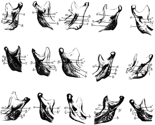
Anatomy Atlases. Image hosted by the site it comes from. Open the original page.
Unerupted Teeth
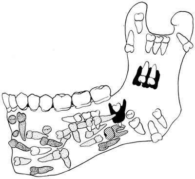
Anatomy Atlases. Image hosted by the site it comes from. Open the original page.
Mental Foramen
Absence and Variations of Mental Foramina
Riferimenti dell’opera originale
Elenco riportato come compare nella fonte, senza normalizzazione dei formati bibliografici.
- Anson, B.J., Ed. (1966) Morris' Human Anatomy, 12th ed., The Blakiston Division, McGraw-Hill Book Co., New York.
- Bradley, O.C. (1902) Two cases of dental anomaly. J. Anat. Physiol. 36:356-367.
- Brocher, J.E.W. (1947) Le processus paracondyleus s. apophyse paramastoid (dans un cas de dysostose mandibulo faciale unilatérale). Radiol. Clin. 16:393-396.
- Dubreuil-Chambardel, L. (1906) Les trous de la symphyse du menton. Assoc. Anatomistes Comptes Rendus 8:14-21.
- Franceschetti, A. (1944) Un syndrome nouveau:la dysotose mandibulo-faciale. Bull. Schweiz. Akad. Med. Wissen. 1(2):60-66. Cited in Excerpta Medica, Sec. 1, Vol. 2, abstract 193, p.98, 1948.
- Gruber, W. (1872) Bemerkungen über das Foramen mentale. Arch. Anat. Physiol. Wissen. Med. 1872:738-745.
- Gruber, W. (1873) über superinfundibulum inframaxillare, den Sulcus mylohyoideus und die beide deckenden knöchernen Brükken. Arch. Anat. Physiol. Wissen. Med. 1873:348-356.
- Gruber, W. (1877) Persistirende unwollständige Theilung des Unterkiefers in zwei Hälften. Arch. Pathol. Anat. Physiol. Klin. Med. 70:137-139.
- Hrdlicka, A. (1941) A lower jaw: Double condyles. Am. J. Phys. Anthropol. 28:75-89.
- Kazanjian, V.H. (1939) Congenital absence of the ramus of the mandible. J. Bone Joint Surg. 21:761-772.
- Keen, J.A. (1945) A study of the angle of the mandible. J. Dent. Res. 24:77-86.
- Rees, L.A. (1954) The structure and function of the mandibular joint. Brit. Dent. J. 96:125-133.
- Santini, A. and M. Land. (1990) A comparison of the positions of the mental foramen in Chinese and British madibles. Acta Anat. 137:208-212.
- Sweet, A.P.S. (1959) Radiodontic study of the mental foramen. Dent. Radiogr. 32:28, 32-33.
- Tero, H.G. and I.R. Telford (1950) An analysis of the variations in the position of the mental foramen. Anat. Rec. 107:61-66.
Anatomical classification
- Systems:
- Skeletal system
Content provenance
Adaptation
Adaptation of «Mandible — Illustrated Encyclopedia of Human Anatomic Variation: Opus I: Muscular System» by Ronald A. Bergman, Adel K. Afifi, Ryosuke Miyauchi. Source: https://www.anatomyatlases.org/AnatomicVariants/SkeletalSystem/Text/Mandible.shtml Original work distributed under the CC BY-NC-SA 4.0 licence (https://creativecommons.org/licenses/by-nc-sa/4.0/deed.it). Adaptation by Redazione AIVA. Changes from the original: Testo originale inglese riprodotto integralmente e riorganizzato nel formato scheda del portale (sintesi, testo, riferimenti), senza modificarne le affermazioni. La traduzione italiana non è ancora stata eseguita, e la pagina lo dichiara. Le figure non sono state copiate: sono collegate alla pagina d’origine. This publication is not approved or endorsed by the authors of the original work.
Licence
This content is distributed under the licence CC BY-NC-SA 4.0. Licences and attribution
